
A groundbreaking investigation by scientists at Scripps Research has unveiled a potentially revolutionary approach to identifying Alzheimer’s disease, moving beyond traditional measures of protein quantity to analyze the intricate structural configurations of proteins circulating in the bloodstream. This innovative methodology, detailed in the February 27, 2026, issue of Nature Aging, reveals that subtle alterations in the way specific plasma proteins are folded can serve as robust indicators of the disease’s presence and progression. The research team successfully differentiated between individuals with normal cognitive function, those experiencing mild cognitive impairment (MCI), and individuals diagnosed with Alzheimer’s disease itself, suggesting a pathway towards earlier diagnosis and intervention.
The scientific community has long recognized the hallmarks of Alzheimer’s disease as the accumulation of amyloid plaques and neurofibrillary tangles within the brain. However, a growing consensus suggests that the disease’s pathology is far more complex, stemming from a systemic breakdown in proteostasis – the biological machinery responsible for ensuring proteins maintain their correct three-dimensional shapes and for clearing away misfolded or damaged protein aggregates. As individuals age, the efficiency of this crucial cellular maintenance system tends to decline, leading to an increased likelihood of proteins adopting aberrant structures. This fundamental principle underpins the Scripps Research team’s hypothesis: if the brain’s proteostasis is compromised by neurodegenerative processes, analogous structural deviations might manifest in the proteins found within the peripheral circulation.
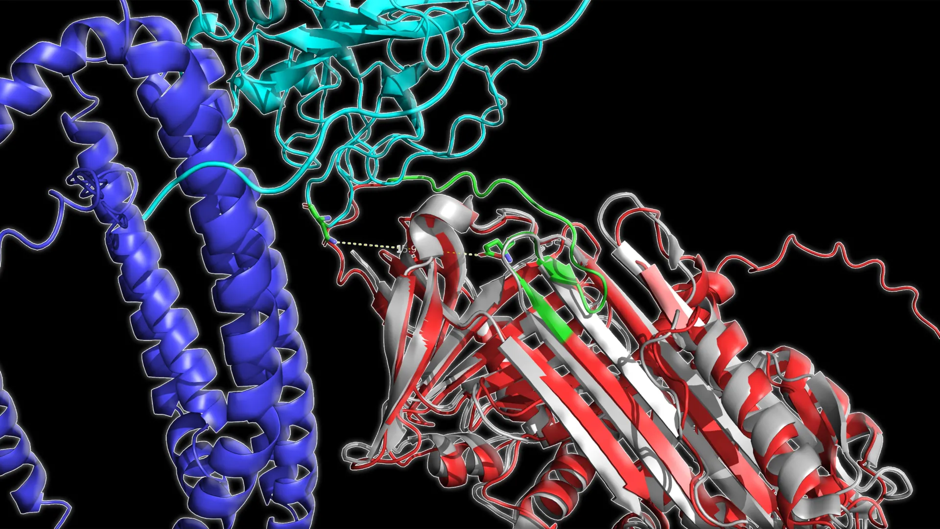
To rigorously test this hypothesis, the researchers meticulously examined plasma samples drawn from a cohort of 520 participants, strategically divided into three distinct clinical categories: cognitively healthy adults, individuals diagnosed with mild cognitive impairment, and patients formally diagnosed with Alzheimer’s disease. The investigation employed sophisticated mass spectrometry techniques, a powerful analytical tool capable of probing the three-dimensional architecture of proteins. Specifically, the scientists assessed the degree of exposure or burial of particular amino acid residues within protein structures, providing a direct measure of their conformational state. This detailed structural information was then subjected to advanced machine learning algorithms, designed to discern complex patterns and identify correlations with the different stages of Alzheimer’s disease.
The analytical findings presented a striking and consistent picture. A discernible pattern emerged: as Alzheimer’s disease advanced, a subset of circulating proteins exhibited a trend towards becoming less structurally "open" or more compact. Crucially, these observed structural modifications proved to be more diagnostically powerful than simple measurements of protein abundance, offering a deeper insight into the underlying biological changes associated with the disease. This emphasis on protein conformation represents a significant departure from conventional diagnostic strategies.
Delving deeper into the data, the research identified three specific plasma proteins whose structural integrity demonstrated the most pronounced correlation with the disease’s trajectory. These key proteins include C1QA, a component of the innate immune system involved in inflammatory signaling; clusterin, a chaperone protein known to assist in protein folding and the clearance of amyloid-beta; and apolipoprotein B, a critical carrier of lipids in the bloodstream that also plays a role in vascular health. The remarkable degree of association observed at specific sites within these proteins was a source of considerable surprise and excitement for the research team.
The ability of these three structurally altered proteins to accurately categorize participants was impressive, achieving an overall diagnostic accuracy of approximately 83% when distinguishing between all three groups. When comparing individuals from two distinct groups, such as healthy controls versus those with MCI, the classification accuracy surged to over 93%, underscoring the robustness of the structural signature. This level of precision suggests a significant leap forward in the potential for non-invasive diagnostic tools.
Further validation of the model’s reliability was demonstrated through its performance on independent cohorts of participants, as well as its ability to track changes over time when analyzing blood samples collected at different temporal intervals. In longitudinal assessments, where samples were taken months apart, the three-protein structural signature maintained its predictive power, accurately reflecting disease status with approximately 86% accuracy and even showing the capacity to indicate shifts in diagnosis over time. Moreover, the derived "structural score" exhibited a strong correlation with established cognitive assessment scores and a more moderate but significant association with observed brain volume reduction, as measured by MRI.
These collective findings strongly indicate that the analysis of protein structure in blood plasma could serve as a valuable adjunct to existing diagnostic methods focused on amyloid and tau biomarkers. By targeting structural changes intrinsically linked to the fundamental biological processes of neurodegeneration, this novel approach holds the potential to refine the identification of disease stages, facilitate more accurate monitoring of disease progression, and provide an objective measure for evaluating the efficacy of therapeutic interventions.
The implications for early intervention are profound, as emphasized by senior author John Yates. "Detecting markers of Alzheimer’s early is absolutely critical to developing effective therapeutics," he stated. "If treatment can start before significant damage has been done, it may be possible to better preserve long-term memory." The prospect of initiating treatment at a stage where neuronal damage is minimized could fundamentally alter the prognosis for individuals affected by this devastating disease.
While the results are highly encouraging, further validation through larger-scale clinical trials with extended follow-up periods will be essential before this blood test can be implemented in routine clinical practice. The research team is also actively exploring the broader applicability of their structural profiling methodology to other complex diseases, including neurodegenerative conditions like Parkinson’s disease and various forms of cancer, suggesting a wide-ranging potential for this innovative technique. The study, titled "Structural signature of plasma proteins classifies the status of Alzheimer’s disease," was co-authored by Ahrum Son, Hyunsoo Kim, and Jolene K. Diedrich from Scripps Research; Heather M. Wilkins, Jeffrey M. Burns, Jill K. Morris, and Russell H. Swerdlow from the University of Kansas Medical Center; and Robert A. Rissman from the University of California San Diego. Funding for this research was provided by the National Institutes of Health through grants RF1AG061846-01, 5R01AG075862, P30AG072973, and P30-AG066530.
About the Author


