
Alzheimer’s disease, a pervasive neurodegenerative condition, currently impacts an estimated 7.2 million individuals aged 65 and older within the United States, as reported by the Alzheimer’s Association. Conventional diagnostic methodologies primarily rely on quantifying the concentrations of two key proteins, amyloid beta and phosphorylated tau, within either blood serum or cerebrospinal fluid. While these established biomarkers have become standard in clinical practice, their capacity to fully encapsulate the earliest molecular alterations that presage the disease’s onset and evolution remains a subject of ongoing scientific inquiry.
A groundbreaking development from researchers at Scripps Research introduces a paradigm shift in Alzheimer’s diagnostics by shifting focus from protein quantity to protein conformation. This innovative blood-based assay investigates the intricate three-dimensional folding patterns of plasma proteins, rather than their mere abundance. The findings, disseminated in the esteemed journal Nature Aging on February 27, 2026, illuminate a significant correlation between distinct structural variations in three specific plasma proteins and an individual’s Alzheimer’s disease status. This sophisticated analytical framework has demonstrated a remarkable ability to accurately differentiate between individuals exhibiting normal cognitive function, those experiencing mild cognitive impairment (MCI), and patients definitively diagnosed with Alzheimer’s. The potential implications of this discovery are profound, paving the way for earlier disease detection and, consequently, more timely therapeutic interventions.
"The genesis of many neurodegenerative disorders is intrinsically tied to aberrant changes in protein architecture," explained senior author John Yates, a distinguished professor at Scripps Research. "Our central hypothesis revolved around the question of whether specific structural modifications in particular proteins could serve as reliable predictive indicators."
The traditional understanding of Alzheimer’s disease has long been anchored to the pathological hallmarks of amyloid plaques and tau tangles that aggregate within the brain parenchyma. However, a growing consensus within the scientific community posits that the disease may represent a more generalized breakdown of proteostasis – the intricate cellular machinery responsible for ensuring proteins achieve and maintain their correct three-dimensional structures, as well as for the efficient clearance of misfolded or damaged protein entities.
As the human aging process unfolds, the efficacy of this vital proteostatic system tends to diminish. This decline renders proteins more susceptible to misfolding during their synthesis or subsequent cellular maintenance. Building upon this foundational principle, the research team hypothesized that if proteostasis is compromised within the central nervous system, analogous structural deviations might manifest in proteins circulating throughout the peripheral bloodstream.
To rigorously test this hypothesis, the research cadre meticulously analyzed plasma samples drawn from a cohort of 520 participants, strategically stratified into three distinct groups: cognitively healthy adults, individuals diagnosed with mild cognitive impairment, and patients formally diagnosed with Alzheimer’s disease.
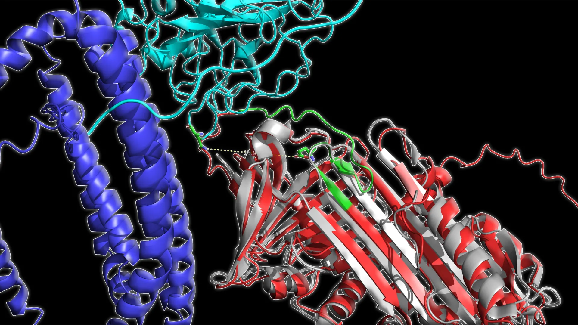
Employing state-of-the-art mass spectrometry, the scientists were able to precisely ascertain the degree of accessibility or sequestration of specific amino acid residues within protein structures. This meticulous analysis provided a quantitative measure of structural alterations. Subsequently, sophisticated machine learning algorithms were deployed to discern discernible patterns within this structural data that bore a discernible relationship to the participant’s disease severity.
The analytical results yielded a compelling and consistent pattern across all participant groups. The study revealed a trend wherein, as Alzheimer’s disease progressed, certain circulating plasma proteins exhibited a reduction in their structural "openness." These observed structural modifications proved to be significantly more informative for delineating the stage of the disease compared to traditional methods that solely assess protein concentration.
Among the vast array of proteins examined, three proteins emerged as exhibiting the most robust association with the progression of Alzheimer’s disease. These critical proteins were identified as C1QA, a key player in immune signaling pathways; clusterin, a protein implicated in both protein folding and the clearance of amyloid aggregates; and apolipoprotein B, a crucial lipoprotein responsible for the transport of fats within the bloodstream and a significant contributor to vascular integrity.
"The magnitude of the correlation we observed was truly astonishing," remarked co-author Casimir Bamberger, a senior scientist at Scripps Research. "It was remarkably unexpected to identify three specific lysine residues, located on three distinct proteins, that demonstrated such a high degree of correlation with the prevailing disease state."
The precise structural modifications identified at these specific sites within the three proteins enabled the researchers to classify participants into their respective diagnostic categories – cognitively normal, MCI, or Alzheimer’s – with an impressive overall accuracy rate of approximately 83%. When direct comparisons were made between two specific groups, such as healthy individuals versus those with MCI, the diagnostic precision escalated to over 93%.
The robustness of this three-protein structural signature was further validated through independent testing on separate participant cohorts. Moreover, the model maintained its reliability when applied to blood samples collected at different time points, extending over several months.
In longitudinal analyses involving repeat blood draws spaced months apart, the developed protein structural panel accurately identified disease status with an average accuracy of approximately 86%. Crucially, it also demonstrated the capacity to reflect shifts in diagnostic classification over time. The calculated structural score exhibited a strong concordance with performance on established cognitive assessments and a more moderate but significant correlation with volumetric measurements of brain atrophy derived from Magnetic Resonance Imaging (MRI).
Collectively, these compelling findings strongly suggest that the analysis of protein structural integrity in blood holds significant promise as a complementary diagnostic tool to existing amyloid and tau-based assays. By concentrating on structural changes that are intrinsically linked to the fundamental biological underpinnings of the disease, this novel methodology may offer enhanced capabilities for precisely identifying disease stages, meticulously monitoring disease progression, and rigorously evaluating the efficacy of therapeutic interventions.
"The early detection of Alzheimer’s disease markers is absolutely paramount for the successful development of effective therapeutic strategies," emphasized Yates. "If treatment can be initiated before substantial and irreversible neurological damage has occurred, there is a far greater potential to preserve long-term cognitive function and memory."
Prior to its widespread adoption in clinical settings, this promising blood-based diagnostic approach necessitates further rigorous validation through larger-scale studies characterized by extended follow-up periods. The research team is also actively exploring the applicability of this structural profiling methodology to other complex diseases, including Parkinson’s disease and various forms of cancer, underscoring the broad potential of this innovative technique.
In addition to the contributions of Yates and Bamberger, the groundbreaking study, titled "Structural signature of plasma proteins classifies the status of Alzheimer’s disease," was co-authored by Ahrum Son, Hyunsoo Kim, and Jolene K. Diedrich from Scripps Research; Heather M. Wilkins, Jeffrey M. Burns, Jill K. Morris, and Russell H. Swerdlow from the University of Kansas Medical Center; and Robert A. Rissman from the University of California San Diego.
The research underpinning this study received crucial financial support from the National Institutes of Health, specifically through grants RF1AG061846-01, 5R01AG075862, P30AG072973, and P30-AG066530.
About the Author


