
Major Depressive Disorder (MDD) presents a profound and pervasive global health challenge, frequently leading to significant disability worldwide. A substantial subset of individuals, estimated at around 30%, experience treatment-resistant depression (TRD), a condition where conventional antidepressant medications fail to elicit a sufficiently positive response. Within this clinical landscape, ketamine has emerged as a promising therapeutic agent, lauded for its remarkably swift antidepressant properties, particularly for those battling TRD. Nevertheless, the precise neural underpinnings of ketamine’s action within the human brain have remained a subject of considerable scientific inquiry, posing an obstacle to the refinement and personalization of this impactful treatment modality.
A pivotal new investigation, disseminated in the esteemed journal Molecular Psychiatry on March 5, 2026, was meticulously designed to demystify this long-standing enigma. Spearheaded by Professor Takuya Takahashi from the Department of Physiology at Yokohama City University Graduate School of Medicine in Japan, the research team employed a sophisticated positron emission tomography (PET) imaging methodology. This cutting-edge technique enabled direct visualization of changes in alpha-amino-3-hydroxy-5-methyl-4-isoxazole propionic acid receptors (AMPARs), a crucial class of proteins instrumental in mediating neuronal communication. These receptors play a vital role in synaptic plasticity and the intricate signaling pathways governed by glutamate, a key neurotransmitter, within individuals undergoing ketamine therapy.
Professor Takahashi articulated the prevailing knowledge gap, stating, "Despite ketamine demonstrating prompt antidepressant effects in individuals diagnosed with treatment-resistant depression, its fundamental molecular mechanism within the human brain has eluded definitive explanation." This uncertainty underscored the imperative for direct observation in living human subjects.
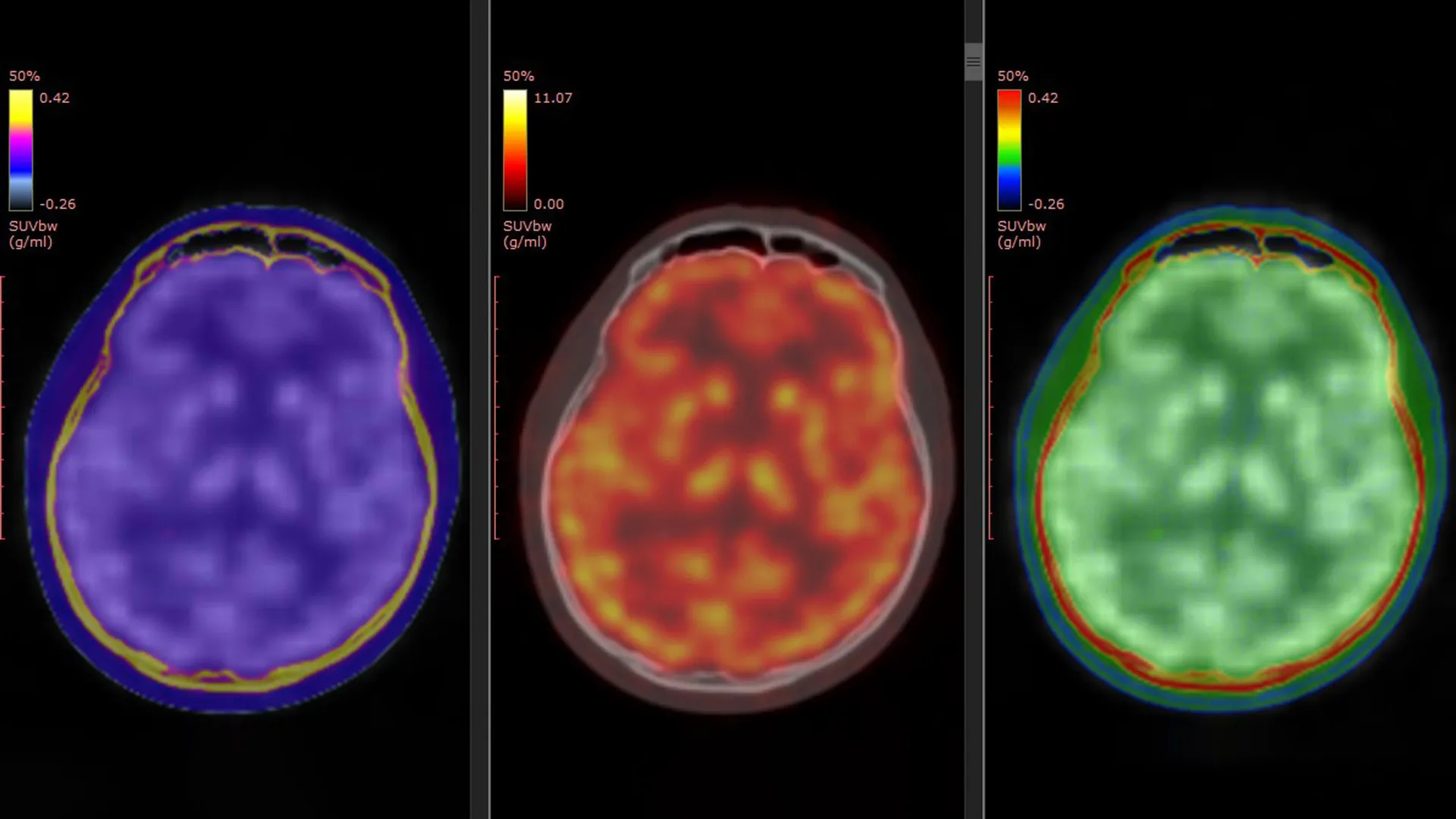
Visualizing Brain Receptors with a Novel PET Tracer
The success of this groundbreaking research hinged upon the application of a specialized PET tracer, designated [¹¹C]K-2, which had been developed by the same research group. This innovative tracer possesses the unique capability to render AMPARs located on the surface of brain cells visible within the living human brain. While prior preclinical studies, conducted in laboratory settings and on animal models, had posited a connection between ketamine’s antidepressant actions and AMPAR activity, this new study offers the first direct empirical substantiation of this process occurring in human subjects.
To facilitate the investigation, the researchers meticulously compiled and analyzed data drawn from three distinct, formally registered clinical trials that had been conducted within Japan. The comprehensive cohort comprised 34 patients formally diagnosed with TRD, alongside 49 healthy individuals who served as a crucial control group for comparative analysis.
Over a two-week treatment period, the patient group received either intravenous ketamine or a placebo administration. Positron emission tomography (PET) brain scans were systematically performed at two key junctures: immediately prior to the commencement of treatment and again following the conclusion of the final infusion. This longitudinal imaging approach provided researchers with a powerful means to meticulously compare and contrast alterations in AMPAR density and distribution within the brain over the course of the intervention.
Region-Specific Brain Changes Linked to Symptom Relief
The analytical findings revealed significant and widespread anomalies in AMPAR density among individuals diagnosed with TRD when contrasted with their healthy counterparts. Importantly, these observed discrepancies were not uniformly distributed across the entire brain but were instead concentrated in specific neural regions.
The administration of ketamine did not induce a homogeneous alteration in receptor levels throughout the cerebral landscape. Instead, the study demonstrated a compelling correlation between improvements in depressive symptomatology and dynamic, region-specific adjustments in AMPAR abundance. Certain cortical areas exhibited an augmentation in receptor density, signifying enhanced signaling capacity. Conversely, other brain regions, particularly those implicated in reward processing, such as the habenula, displayed a reduction in AMPAR levels. These precisely localized shifts in AMPAR distribution were found to be strongly associated with the degree of symptomatic relief experienced by the patients.
Professor Takahashi further elucidated this critical observation: "Ketamine’s antidepressant impact in patients suffering from TRD is facilitated by dynamic modifications in AMPARs within the living human brain. Utilizing the novel PET tracer, [¹¹C]K-2, we were able to visualize the specific ways in which ketamine influences AMPAR distribution across distinct brain regions and, crucially, how these observed alterations correlate with amelioration of depressive symptoms." These findings represent a direct translation of previously identified mechanisms from animal studies into observable human clinical effects, thereby bridging a significant gap in our understanding.
Potential Biomarker for Predicting Treatment Response
Beyond their contribution to elucidating the neurobiological mechanisms of ketamine, these findings hold significant promise for practical clinical application. The capacity to image AMPARs using PET technology could potentially evolve into a valuable biomarker. Such a biomarker could empower clinicians to more accurately assess and predict an individual patient’s likelihood of responding favorably to ketamine treatment.
Given that a considerable number of patients do not achieve adequate relief from standard antidepressant therapies, the identification of reliable biological indicators for predicting treatment outcomes remains a paramount objective within the field of mental healthcare. This research offers a tangible step toward achieving that goal.
Toward More Personalized Depression Treatments
By providing scientists with the unprecedented ability to directly observe AMPAR activity within the living human brain, this research significantly narrows the long-standing chasm between foundational laboratory discoveries and applied clinical psychiatry. The results firmly establish AMPAR modulation as a central mechanistic pathway underpinning ketamine’s rapid antidepressant effects. Furthermore, they strongly suggest that AMPAR-focused PET imaging could serve as a powerful tool to guide the development and implementation of increasingly personalized therapeutic strategies in the future.
Ultimately, this pioneering work has the potential to accelerate the creation of more precise and effective interventions for individuals grappling with the debilitating effects of treatment-resistant depression, thereby enhancing their quality of life and fostering a more optimistic outlook for their therapeutic journey.
This research was made possible through the generous support of various governmental and foundational entities, including the Ministry of Education, Culture, Sports, Science and Technology (under the Special Coordination Funds for Promoting Science and Technology); the Japan Agency for Medical Research and Development (AMED), providing grant numbers JP18dm0207023, JP19dm0207072, JP24wm0625304, JP25gm7010019, and JP20dm0107124; the Japan Society for the Promotion of Science KAKENHI, with grant numbers 22H03001, 20H00549, 20H05922, 23K10432, 19H03587, 20K20603, 22K15793, and 21K07508; the Takeda Science Foundation; the Keio Next-Generation Research Project Program; the SENSHIN Medical Research Foundation; and the Japan Research Foundation for Clinical Pharmacology.
About the Author


