
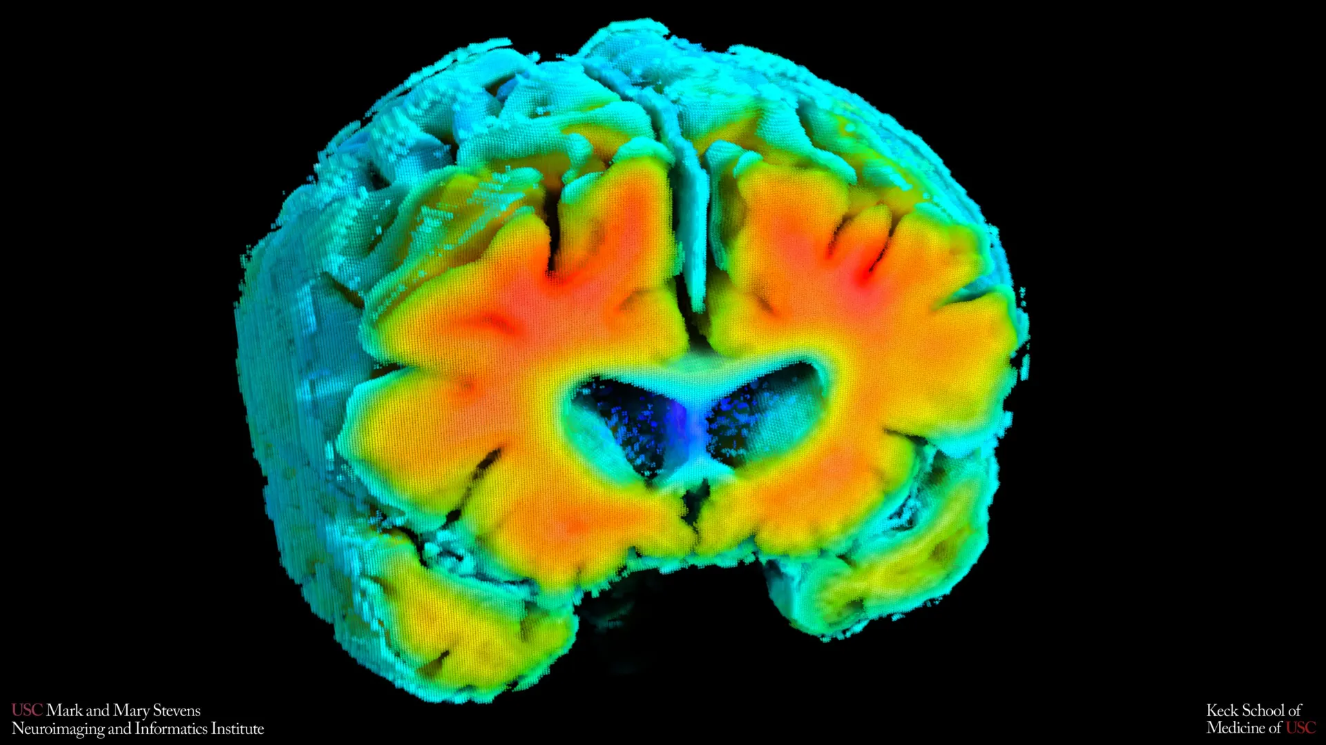
New scientific inquiry spearheaded by researchers at the Mark and Mary Stevens Neuroimaging and Informatics Institute (Stevens INI), an arm of the Keck School of Medicine of USC, posits a significant correlation between subtle alterations in cerebral circulation and the foundational stages of Alzheimer’s disease. This groundbreaking research, detailed in the latest issue of Alzheimer’s and Dementia: The Journal of the Alzheimer’s Association, suggests that the efficient delivery of oxygen to brain tissue and the smooth movement of blood throughout the cranial vasculature may play a more pivotal role in the disease’s genesis than previously understood, potentially serving as an early warning signal long before cognitive symptoms manifest.
The investigative team meticulously analyzed data from a cohort of older adults, encompassing individuals with and without diagnosed cognitive impairments. Employing a suite of non-invasive methodologies, the researchers quantified key aspects of brain blood flow and oxygenation. Their findings illuminated a discernible connection between these physiological markers and established hallmarks of Alzheimer’s, such as the pathological accumulation of amyloid plaques—protein fragments that aggregate in the brain—and the reduction in volume of the hippocampus, a brain structure critically involved in memory formation and retrieval. The implications are substantial: the integrity of the brain’s vascular network appears to exert an early influence on the cascade of events leading to Alzheimer’s, offering a promising avenue for identifying at-risk individuals at an earlier, more treatable stage.
Amaryllis A. Tsiknia, the lead author of the study and a doctoral candidate at USC, emphasized the evolving understanding of Alzheimer’s pathology. While amyloid and tau proteins have traditionally been regarded as the primary culprits driving neurodegeneration in Alzheimer’s, Tsiknia highlighted the indispensable role of vascular function. "Our findings underscore that when the brain’s vascular system operates with the resilience and adaptability characteristic of healthy aging, we concurrently observe brain structural and biochemical profiles that are associated with superior cognitive function," she stated, pointing to a paradigm shift in how the disease is conceptualized.
To meticulously assess these delicate circulatory shifts, the research utilized two straightforward, painless techniques adaptable for individuals at rest. Transcranial Doppler ultrasound provided a means to measure the velocity of blood flow through the brain’s principal arteries, offering insights into the speed at which blood navigates these vital channels. Concurrently, near-infrared spectroscopy (NIRS) was employed to gauge the efficacy with which oxygenated blood reached the superficial layers of the cerebral cortex, indicating the efficiency of oxygen transfer to neural tissue.
Sophisticated mathematical algorithms were then applied to synthesize the data derived from these imaging modalities. This advanced computational approach yielded composite indicators of cerebrovascular function. These integrated metrics provided a holistic assessment of the brain’s capacity to dynamically regulate blood flow and oxygen supply in response to physiological variations, such as fluctuations in blood pressure and carbon dioxide levels, which are normal, ongoing processes within the body. This dynamic regulation is crucial for maintaining neuronal health and optimal brain function.
The study revealed a compelling pattern: participants whose cerebrovascular function indicators more closely mirrored those observed in individuals with unimpaired cognition exhibited lower levels of amyloid deposition and possessed larger hippocampal volumes. Both of these attributes are independently recognized as protective factors against the development of Alzheimer’s disease. This suggests a fundamental link between vascular health and the core pathological features of Alzheimer’s.
Dr. Meredith N. Braskie, a senior author on the study and an assistant professor of neurology at Keck School of Medicine, commented on the significance of these vascular measures. "These vascular metrics are capturing something profoundly meaningful about the overall health of the brain," she observed. "They appear to correlate strongly with findings typically observed through Magnetic Resonance Imaging (MRI) and Positron Emission Tomography (PET) scans, which are the current gold standards for investigating Alzheimer’s disease. This provides critical context for understanding the intricate relationship between vascular well-being and the established neuroimaging markers of Alzheimer’s risk."
Further reinforcing the study’s conclusions, individuals who had been diagnosed with mild cognitive impairment (MCI) or dementia displayed demonstrably weaker cerebrovascular function when compared to their cognitively normal counterparts. This observation lends considerable weight to the hypothesis that a decline in the health of the brain’s blood vessels is not an isolated issue but rather an integral component of the broader pathological continuum associated with Alzheimer’s disease.
Dr. Arthur W. Toga, director of the Stevens INI, articulated the growing consensus within the scientific community. "These findings contribute to a mounting body of evidence suggesting that Alzheimer’s disease involves significant vascular contributions, acting in concert with, and potentially preceding, the classic neurodegenerative changes," he stated. "Elucidating the complex interplay between cerebral blood flow, oxygen regulation, amyloid pathology, and structural brain alterations opens up exciting new avenues for the development of earlier diagnostic tools and potentially novel preventive strategies."
The practical implications of these non-invasive assessment methods are particularly noteworthy. When juxtaposed with the more resource-intensive and complex procedures of MRI and PET imaging, the techniques employed in this study offer distinct advantages. They are considerably less expensive to implement, more readily accessible, and impose a lower burden on participants. Crucially, they do not necessitate the administration of contrast agents, exposure to ionizing radiation, or the need for individuals to perform demanding cognitive tasks. This inherent simplicity and ease of use position these methods as potentially invaluable for large-scale population screening initiatives or for individuals for whom more invasive or strenuous brain imaging modalities are not feasible or appropriate.
However, the authors of the study exercise appropriate scientific caution, acknowledging that their findings represent a cross-sectional snapshot in time. This means that while associations have been identified, the research as presented does not definitively establish a cause-and-effect relationship. To address this, longitudinal studies are currently underway, involving the ongoing monitoring of participants. The aim of these long-term investigations is to determine whether observed shifts in these vascular parameters can reliably predict future cognitive decline or gauge an individual’s response to therapeutic interventions.
"Should our ongoing research confirm that these vascular signals can be tracked prospectively over time, we may gain the unprecedented ability to identify individuals at heightened risk for Alzheimer’s disease at a significantly earlier juncture," Tsiknia explained. "Furthermore, this could enable us to rigorously test whether interventions aimed at improving vascular health can effectively mitigate or even reverse the brain changes associated with Alzheimer’s pathology."
The research team responsible for this insightful study included, in addition to Tsiknia and Braskie, Peter S. Conti, Rebecca J. Lepping, Brendan J. Kelley, Rong Zhang, Sandra A. Billinger, Helena C. Chui, and Vasilis Z. Marmarelis. Funding for this critical work was generously provided by the Office of The Director, National Institutes of Health, under Award Number S10OD032285, and by the National Institute on Aging through grant R01AG058162. This collaborative effort represents a significant step forward in understanding the multifaceted origins of Alzheimer’s disease and holds considerable promise for future diagnostic and therapeutic advancements.
About the Author


