
Brainstem’s "Blue Place" Orchestrates Targeted Neural Communication, Redefining Neurotransmitter Signaling.
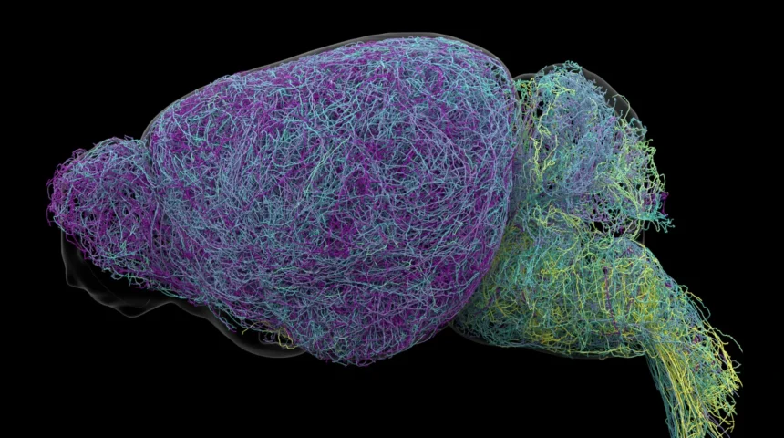
Residing in the core of the brainstem, a remarkably compact cluster of nerve cells, historically known as the locus coeruleus (LC) from the Latin for "blue place," exerts a disproportionately influential role in the brain’s overall operational capacity. Despite comprising a minuscule fraction of the brain’s total neuronal population, this region is the primary source of norepinephrine (NE), a crucial chemical messenger that profoundly shapes cognitive functions such as attention and learning, influences autonomic processes like heart rate, and modulates the body’s intricate responses to stress and moments of heightened arousal. For decades, the prevailing scientific consensus posited that the LC functioned akin to a broadcast tower, disseminating a singular, undifferentiated norepinephrine signal throughout the vast neural landscape. However, groundbreaking research recently published in the esteemed journal Nature has systematically dismantled this long-held assumption, revealing a far more nuanced and sophisticated communication paradigm.
Employing a sophisticated array of advanced methodologies, including cutting-edge brain imaging techniques, precise recordings of neural electrical activity, advanced genetic manipulation strategies, and meticulously designed behavioral experiments conducted with rodent models, a team of investigators at the Allen Institute has unveiled compelling evidence that the LC operates not as a monolithic broadcaster but rather through a network of multiple, distinct neural pathways. This revolutionary discovery posits that instead of emitting a generalized signal, the LC delivers highly specific messages to designated neural destinations, with different subpopulations of neurons exhibiting differential responses to varying external stimuli and internal experiences. This fundamental shift in understanding reconfigures our perception of how this critical neuromodulatory system operates.
Unveiling a Sophisticated Neural Network Capable of Precision Messaging
Through the meticulous tracing of axonal projections originating from LC neurons, the researchers have definitively established that discrete groups of these cells establish connections with particular regions of the nervous system. These identified pathways exert direct control over the precise locations where norepinephrine is released, thereby constructing a communication architecture that is demonstrably more organized and targeted than previously conceptualized. The anatomical organization within the LC further underscores this refined understanding. Neurons situated within the dorsal, or upper, aspect of the LC predominantly project their signals to the cerebral cortex and forebrain, regions critically involved in higher-order cognitive functions. Conversely, neurons located in the ventral, or lower, portion of the LC primarily transmit their signals to the brainstem and the spinal cord, areas responsible for regulating more fundamental bodily functions and motor control. This spatial segregation of neural connections empowers distinct subregions of the LC to exert influence over entirely separate neural circuits and, consequently, distinct brain functions.
Dr. Karel Svoboda, Director of Neural Dynamics at the Allen Institute and a senior author on the study, eloquently summarized this paradigm shift by stating, "The findings suggest the brain’s norepinephrine system is far more like a targeted postal network than a foghorn." This analogy effectively conveys the transition from a perception of indiscriminate broadcasting to one of precise, directed delivery, akin to a sophisticated messaging system.
Profound Implications for Understanding and Treating Neurological and Psychiatric Disorders
The elucidation of this intricate signaling network holds profound potential for transforming the therapeutic landscape for a spectrum of neurological and mental health conditions. Norepinephrine’s pivotal role in modulating attention, mood, and emotional regulation positions it as a critical target for pharmacological interventions employed in the treatment of prevalent disorders such as depression, anxiety, and Attention-Deficit/Hyperactivity Disorder (ADHD). Furthermore, neurons within the LC are among the earliest to exhibit pathological deterioration in neurodegenerative conditions like Alzheimer’s disease, suggesting that these novel findings could offer crucial insights into the underlying mechanisms of these devastating illnesses and pave the way for the development of more effective treatments.
Current therapeutic strategies that aim to modulate norepinephrine levels often exert their effects across multiple, widespread brain circuits. While this broad action can yield beneficial outcomes in certain neural pathways, it can also precipitate undesirable side effects in others due to the lack of specificity. A comprehensive and detailed map of the LC’s specialized projection pathways could empower scientists to engineer therapies that exhibit remarkable precision, selectively targeting the neural circuits implicated in specific symptom constellations without adversely affecting other vital brain functions. This advancement promises a future where treatments are not only more efficacious but also significantly safer and better tolerated.
This groundbreaking research was generously supported by the National Institutes of Health’s Brain Research Through Advancing Innovative Neurotechnologies® (BRAIN) Initiative, a testament to the national commitment to unraveling the complexities of the brain.
Identification of Distinct Neuronal Roles in Learning and Environmental Engagement
Beyond delineating the anatomical pathways, the research has also uncovered a critical functional divergence between different populations of LC neurons. Specifically, neurons within the dorsal LC that project to the cerebral cortex demonstrated heightened activity during learning paradigms. In experimental settings where mice were engaged in decision-making tasks and subsequently adjusted their behavioral choices following an unfavorable outcome, these specific neurons exhibited a marked increase in their firing rate. This observation strongly suggests their direct involvement in the process of adaptive learning and behavioral modification in response to feedback.
In contrast, neurons located in the ventral LC exhibited a distinct pattern of activity. Their firing rates were observed to increase transiently prior to instances where mice actively ignored cues that signaled the potential for an available reward. This behavioral pattern indicates that these ventral LC neurons play a crucial role in regulating the organism’s propensity to engage with its environment and respond to salient stimuli, effectively acting as gatekeepers of behavioral initiation.
Dr. Svoboda further elaborated on these functional distinctions: "What emerged was a clear map: neurons in the dorsal LC that send signals upward to the cortex are involved in learning, whereas neurons in the ventral region projecting downward to the brainstem and spinal cord govern whether animals engage with their environment at all." This encapsulates the functional dichotomy observed. The study also provided compelling evidence that these anatomical and functional differences are mirrored by distinct patterns of gene expression, reinforcing the notion of highly specialized neuronal subpopulations within the LC. This indicates that the differentiation extends beyond mere anatomical location and connectivity to encompass fundamental molecular mechanisms that underpin their unique roles.
Discovery of Exceptionally Long Neuronal Axons
The comprehensive nature of this investigation involved an exhaustive examination of the mouse brain, utilizing whole-brain imaging to analyze nearly 35,000 individual neurons. This was complemented by an in-depth analysis of the genetic profiles of approximately 400,000 cells, and the meticulous reconstruction of the complete morphological structures of several exceptionally intricate nerve cells. During this extensive data acquisition and analysis process, the researchers stumbled upon another remarkable and unexpected finding: LC neurons possess extraordinarily elongated axons, the projections that transmit neural signals from one neuron to another.
The research team found that the axons of LC neurons measured an average of approximately 35 centimeters in length. In a particularly striking case, one reconstructed axon reached a staggering total length of 70.32 centimeters, classifying it as one of the longest single neurons ever documented in a mouse. Dr. Jeremiah Cohen, a scientist at the Allen Institute and co-author of the study, remarked, "This neuron, like many others that we studied, supplies NE to a very large volume of the cerebral cortex. For the brain, this is highly unusual. Most neurons are more specific in their targets. But this neuron doesn’t release NE everywhere. It ignores the cerebellum, brainstem, and spinal cord." This observation, while highlighting the remarkable physical dimensions of these neurons, paradoxically reinforces the central discovery: despite their extensive reach, these neurons do not indiscriminately distribute norepinephrine throughout the entire brain. Instead, their connectivity remains remarkably selective, underscoring the existence of precisely organized signaling pathways within the LC.
A Paradigm Shift in Understanding Neural Learning Mechanisms
The research further illuminated intriguing parallels between the norepinephrine system and the brain’s dopamine signaling system, another key neuromodulatory pathway. Dopamine, a critical chemical messenger, transmits signals to the basal ganglia, a brain region intimately involved in the formation of habits and reward-based learning. Scientists have long hypothesized that dopamine and norepinephrine likely collaborate in a synergistic manner to facilitate learning processes. This collaborative effort may enable the brain to concurrently acquire and consolidate multiple complex and abstract concepts, contributing to a more comprehensive and flexible understanding of the world.
By meticulously dissecting the intricate connectivity patterns, elucidating the specialized functional roles, and characterizing the unusual physical architecture of neurons within the locus coeruleus, this comprehensive study has significantly advanced our understanding of how the animal brain communicates, learns, and interacts with its environment. Although the findings are derived from experiments conducted with rodent models, their implications are expected to extend to human brain function, offering a more refined framework for comprehending human cognition and paving the way for the development of targeted therapeutic interventions that precisely modulate specific neural circuits, rather than broadly impacting entire signaling systems. This promises a future of more precise and effective neurological and psychiatric treatments.

Related News