A groundbreaking development in regenerative medicine from a Stanford Medicine-led research team suggests a novel approach to address age-related cartilage degeneration and the debilitating progression of osteoarthritis. Scientists have identified a specific protein, whose concentrations escalate with advancing age, as a key target for intervention, demonstrating success in restoring joint cartilage in preclinical models and human tissue samples. This innovative strategy offers a potential paradigm shift from merely managing symptoms to actively regenerating the vital cushioning tissue within joints, holding profound implications for millions afflicted by chronic joint pain and limited mobility.
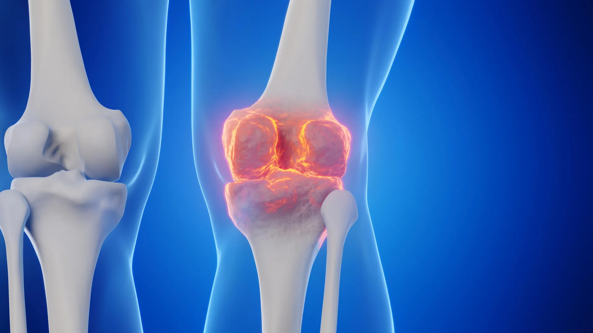
Osteoarthritis (OA), a prevalent degenerative joint condition, is characterized by the gradual erosion of cartilage, the flexible connective tissue that covers the ends of bones in a joint. This deterioration leads to pain, inflammation, swelling, and progressively restricted joint movement. Affecting approximately one in five adults across the United States, OA represents a substantial public health burden, with annual direct healthcare expenditures estimated at around $65 billion. Current therapeutic options primarily focus on alleviating discomfort and preserving joint function through physical therapy, pain medications, and, in advanced stages, surgical interventions like total joint replacement. Crucially, there remains no pharmacological agent capable of reliably halting or reversing the underlying pathological process of cartilage loss.
The recent investigation centered on an enzyme designated 15-PGDH (15-hydroxyprostaglandin dehydrogenase). This enzyme has been categorized by the researchers as a "gerozyme," a term coined to describe enzymes that become increasingly abundant with biological aging and contribute to the functional decline of various tissues throughout the body. Previous studies conducted by the same research group had established 15-PGDH’s multifaceted role as a critical regulator of the aging process across multiple tissue types. For instance, inhibiting this protein with a small molecule compound was shown to augment muscle mass and enhance endurance in older mice. Conversely, elevating 15-PGDH levels in younger animals resulted in muscle atrophy and weakening, underscoring its pivotal role in tissue maintenance and function. Beyond musculoskeletal tissue, the enzyme has also been implicated in the regenerative capacities of bone, nerve, and blood cells.
A central premise of many regenerative processes in diverse tissues involves the proliferation and differentiation of tissue-specific stem cells into specialized cell types. However, the current research revealed a distinct mechanism at play in cartilage regeneration. Instead of recruiting or activating stem cell populations, the findings indicated that existing cartilage cells, known as chondrocytes, exhibited a remarkable capacity for plasticity. These chondrocytes underwent a reprogramming of their gene expression profiles, effectively reverting to a more youthful, regenerative phenotype. Dr. Helen Blau, a professor of microbiology and immunology and director of the Baxter Laboratory for Stem Cell Biology, emphasized the novelty of this discovery, stating, "This represents a novel pathway for regenerating adult tissue, bearing significant clinical promise for addressing arthritis arising from either aging or injury. Our initial hypothesis anticipated the involvement of stem cells, but their absence in this mechanism is truly exciting." Dr. Blau, alongside Dr. Nidhi Bhutani, an associate professor of orthopaedic surgery, served as senior authors on the research, which was featured in the prestigious journal Science. Dr. Mamta Singla, an instructor of orthopedic surgery, and Dr. Yu Xin (Will) Wang, a former postdoctoral scholar now an assistant professor at the Sanford Burnham Institute in San Diego, were lead authors of the study.
Dr. Bhutani underscored the critical need for effective interventions: "Millions of individuals experience joint discomfort and swelling as they age. This represents a substantial, unmet medical challenge. Until now, no therapeutic agent has directly targeted the root cause of cartilage degradation. Yet, this gerozyme inhibitor instigated a profound restoration of cartilage, surpassing the effects reported for any other drug or treatment strategy."
To comprehend the specific type of cartilage involved, it is important to differentiate between the three primary forms found in the human body, each adapted for distinct physiological roles. Elastic cartilage, characterized by its flexibility, is found in structures such as the outer ear. Fibrocartilage, known for its robust shock-absorbing properties, is present in areas like the spinal discs. Hyaline cartilage, the smoothest and most slippery variety, enables low-friction movement between bones within synovial joints, including the ankles, hips, shoulders, and certain parts of the knee. Within these joints, hyaline cartilage is referred to as articular cartilage, and it is precisely this type that is most frequently compromised in osteoarthritis.
The onset of osteoarthritis can be triggered by chronic stress from aging, traumatic injury, or conditions like obesity. In affected joints, chondrocytes begin to secrete inflammatory molecules while simultaneously breaking down collagen, a structural protein critical for cartilage strength and integrity. As collagen diminishes, the articular cartilage thins and softens. The accompanying inflammation exacerbates swelling and pain, culminating in the characteristic symptoms of OA. A significant challenge in treating OA stems from the inherently limited capacity of articular cartilage for self-repair. While some stem or progenitor cells capable of forming cartilage have been identified within bone, similar cell populations have not been successfully located within the articular cartilage itself, contributing to its poor regenerative potential.
Earlier investigations from Dr. Blau’s laboratory had revealed that prostaglandin E2 (PGE2) plays a crucial role in the proper functioning of muscle stem cells. The enzyme 15-PGDH, in turn, is responsible for the catabolism (breakdown) of PGE2. Consequently, either inhibiting 15-PGDH or directly elevating PGE2 levels had previously been shown to support regenerative processes in damaged muscle, nerve, bone, colon, liver, and blood cells in young mice. These prior findings prompted a critical inquiry by Blau, Bhutani, and their collaborators: Could this same biochemical pathway contribute to the age-related deterioration of cartilage or its inability to recover following injury?
To explore this hypothesis, the researchers compared the levels of 15-PGDH in knee cartilage extracted from young and aged mice. They observed a significant increase, roughly a doubling, of the gerozyme’s concentration with advancing age. Subsequently, older animals were administered a small molecule compound specifically engineered to inhibit 15-PGDH. In one experimental setup, the compound was injected systemically into the abdomen, allowing for widespread distribution throughout the body. In a parallel experiment, the inhibitor was delivered directly into the knee joint. Both methods yielded compelling outcomes.
Articular cartilage in the knees of the older, untreated mice was noticeably thinner and exhibited diminished functionality compared to that of younger animals. However, following treatment with the 15-PGDH inhibitor, the cartilage surface throughout the joint became considerably thicker. Importantly, the newly formed tissue was confirmed to be hyaline cartilage, the smooth articular cartilage essential for healthy joint articulation, rather than the less suitable fibrocartilage. Dr. Bhutani expressed surprise at the extent of the regeneration, remarking, "The degree of cartilage regeneration observed in aged mice was truly unexpected. The effect was quite remarkable."
The research team further investigated the therapeutic potential of the treatment in the context of traumatic knee injuries. Tears to the anterior cruciate ligament (ACL) are particularly common in sports requiring sudden pivots, abrupt stops, or jumping maneuvers, such as soccer, basketball, and skiing. While surgical intervention can repair the torn ligament, addressing the immediate injury does not consistently prevent long-term joint damage. A significant proportion, approximately 50%, of individuals who sustain ACL injuries will eventually develop osteoarthritis in the affected joint within about 15 years.
In the mouse injury experiments, the gerozyme inhibitor was administered twice weekly for a period of four weeks following injury. This treatment dramatically lowered the probability of the animals developing post-traumatic osteoarthritis. In contrast, mice receiving a control drug displayed 15-PGDH levels twice as high as those in uninjured animals and developed clear signs of osteoarthritis within four weeks. Furthermore, the treated mice exhibited more natural movement patterns and placed a greater proportion of their body weight on the paw corresponding to the injured limb compared to their untreated counterparts. Dr. Blau commented on an interesting aspect of the findings: "While prostaglandin E2 has historically been associated with inflammation and pain, this research demonstrates that, within physiological ranges, modest elevations in prostaglandin E2 can actually promote regeneration."
To elucidate the cellular and molecular events occurring within the joint, the investigators conducted a detailed analysis of chondrocytes from young and old mice. Older cartilage cells exhibited heightened activity in genes linked to inflammation and the undesirable transformation of hyaline cartilage into bone. Concurrently, genes crucial for normal cartilage development displayed reduced activity. The therapeutic intervention effectively recalibrated this genetic balance. A specific subpopulation of old chondrocytes, which produced 15-PGDH and expressed genes associated with cartilage degradation, saw its prevalence decrease from 8% to 3% of the total cell count. Another group of cells, which did not produce 15-PGDH but expressed genes indicative of fibrocartilage formation, also diminished from 16% to 8%. Conversely, a third population of chondrocytes demonstrated a sharp increase in representation, rising from 22% to 42%. These cells did not produce 15-PGDH and expressed genes essential for forming hyaline cartilage and maintaining the extracellular matrix, the intricate network of proteins and other molecules surrounding cells that provides structural support and mechanical properties to tissues. In cartilage, the extracellular matrix is particularly vital for enabling the tissue to withstand compressive forces while preserving the smooth surface required for seamless joint articulation. Overall, the treatment appeared to drive cartilage towards a biologically rejuvenated state without necessitating the recruitment of external stem or progenitor cells.
Crucially, the researchers extended their investigation to human tissue. They examined cartilage samples obtained from individuals with osteoarthritis undergoing total knee replacement surgery. Following a single week of treatment with the 15-PGDH inhibitor, the human tissue demonstrated a reduction in chondrocytes expressing 15-PGDH. Activity in genes associated with cartilage degradation and fibrocartilage formation also declined when compared to untreated tissue samples. Most significantly, the treated human cartilage samples commenced a process of articular cartilage regeneration.
Dr. Bhutani reflected on the profound implications of these observations: "The underlying mechanism is quite striking and has genuinely transformed our understanding of how tissue regeneration can occur. It is evident that a substantial pool of pre-existing cells within the cartilage are altering their gene expression patterns. By targeting these cells for regeneration, we may unlock an opportunity to achieve a more significant clinical impact overall."
While these findings represent highly promising early evidence, it is important to note that the studies in mice and ex vivo human tissue do not yet definitively establish that this treatment can regenerate cartilage or prevent osteoarthritis in human patients in vivo. Rigorous clinical trials specifically designed to assess cartilage regeneration will be indispensable to ascertain the safety and efficacy of this approach for patients. Encouragingly, an oral formulation of a 15-PGDH inhibitor has already progressed to clinical testing for a different age-related condition: muscle weakness. Dr. Blau confirmed, "Phase 1 clinical trials of a 15-PGDH inhibitor for muscle weakness have demonstrated its safety and activity in healthy volunteers. Our aspiration is that a similar trial will soon be initiated to evaluate its effects in cartilage regeneration. We are incredibly enthusiastic about this potential breakthrough. The prospect of regenerating existing cartilage and potentially avoiding joint replacement surgery is truly transformative."
Researchers from the Sanford Burnham Prebys Medical Discovery Institute also contributed to this significant work. The study received funding from multiple sources, including the National Institutes of Health (grants R01AR070864, R01AR077530, R01AG069858, and R00NS120278), the Baxter Foundation for Stem Cell Biology, the Li Ka Shing Foundation, the Stanford Cardiovascular Institute, the Milky Way Research Foundation, the Canadian Institutes of Health Research, a Stanford Translational Research and Applied Medicine Pilot grant, a GlaxoSmithKline Sir James Black Postdoctoral Fellowship, and a Stanford Dean’s Postdoctoral Fellowship. It is also noted that Drs. Blau, Bhutani, and other coauthors hold inventor status on patent applications filed by Stanford University concerning 15-PGDH inhibition for cartilage and tissue rejuvenation, which are licensed to Epirium Bio. Dr. Blau is a cofounder of Myoforte/Epirium and maintains equity and stock options in the company, a standard disclosure in scientific reporting.
About the Author