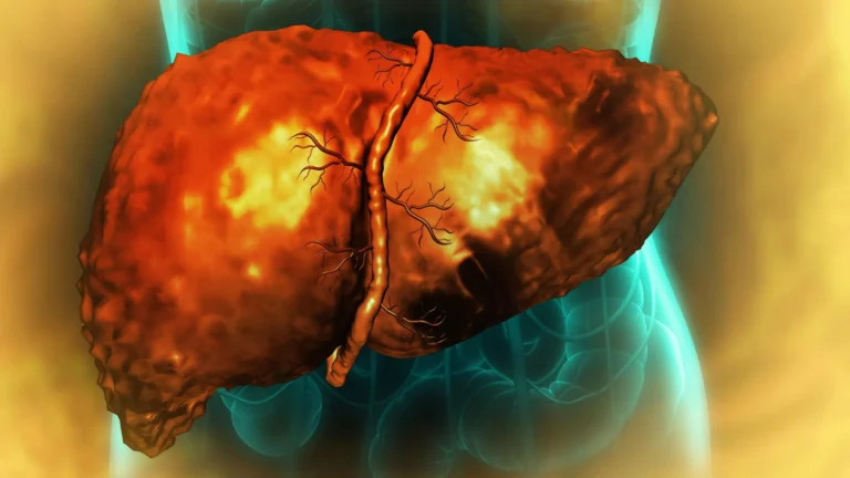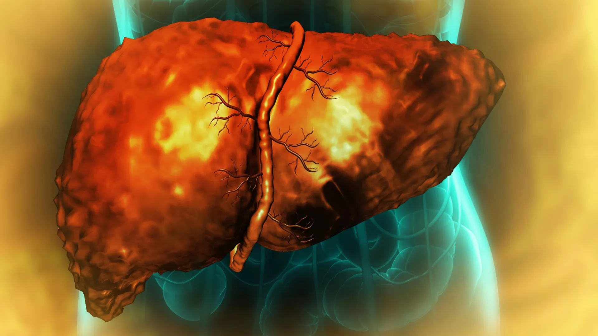
The human liver, a vital organ with over 500 critical functions, possesses a remarkable and nearly unparalleled capacity for self-repair and regeneration following injury. This inherent ability allows it to bounce back from significant damage, distinguishing it from most other major organs. However, chronic excessive alcohol consumption presents a profound challenge to this regenerative prowess, often leading to severe and irreversible conditions. A groundbreaking interdisciplinary investigation has now illuminated a fundamental molecular mechanism through which alcohol-induced damage can permanently impair the liver’s ability to heal itself, even long after an individual ceases drinking. The findings suggest that hepatic cells become locked in an anomalous, non-functional transitional state, unable to complete their natural restorative cycle.
This pivotal research, conducted collaboratively by scientists from the University of Illinois Urbana-Champaign, Duke University, and the Chan Zuckerberg Biohub Chicago, pinpoints chronic inflammation as a primary driver of this cellular stasis. Specifically, the study reveals that inflammatory signals disrupt RNA splicing, an intricate cellular process essential for translating genetic blueprints into functional proteins. Published in the prestigious journal Nature Communications, these insights offer a beacon of hope, potentially paving the way for novel diagnostic tools and therapeutic interventions for severe alcohol-associated liver disease (ALD), a global health crisis.
Alcohol-associated liver disease stands as a leading cause of liver-related mortality worldwide, contributing to approximately three million deaths annually. It encompasses a spectrum of conditions, from fatty liver (steatosis) to alcohol-associated hepatitis (AH) and, ultimately, cirrhosis, a stage characterized by irreversible scarring and impaired liver function. In its advanced forms, ALD often necessitates liver transplantation, a complex procedure with significant limitations, including donor organ scarcity, lifelong immunosuppression, and considerable costs. Understanding the underlying mechanisms that perpetuate liver damage despite alcohol abstinence is therefore critical for developing more accessible and effective treatments.
Historically, medical science has grappled with the perplexing observation that many patients with ALD, particularly those with alcohol-associated hepatitis and cirrhosis, experience progressive liver deterioration even after discontinuing alcohol consumption. Professor Auinash Kalsotra, a biochemistry expert at the University of Illinois Urbana-Champaign and co-lead author of the study, articulated this enigma: "We understood that the liver ceases to function and regenerate in these patients, even post-abstinence, but the precise reasons remained elusive." He underscored the urgency, noting, "Transplantation remains the sole life-saving option at the stage of liver failure. If we could decipher why these livers are failing, we might be able to intervene earlier and more effectively."
The research team, including Professor Anna Mae Diehl from Duke University School of Medicine, has dedicated years to unraveling the molecular intricacies of liver regeneration. Their previous work established that during the repair process, mature liver cells undergo a temporary reprogramming of their gene expression profiles. This involves a crucial step where specialized adult hepatocytes temporarily revert to a more primitive, fetal-like progenitor state. In this less specialized condition, these cells gain the ability to divide and proliferate, thereby generating new tissue to replace damaged areas. Following this proliferative phase, these progenitor-like cells typically mature once again, differentiating into fully functional adult liver cells, thus completing the regenerative cycle and restoring normal hepatic architecture and function. The current study aimed to investigate what disrupts this finely tuned regenerative cascade in the context of alcohol-associated liver disease.
To address this question, the researchers meticulously compared healthy liver tissue samples with those obtained from individuals diagnosed with alcohol-associated hepatitis or cirrhosis. These diseased samples were procured from Johns Hopkins University Hospital through an initiative supported by the National Institute on Alcohol Abuse and Alcoholism, part of the National Institutes of Health, ensuring high-quality, clinically relevant specimens. A consistent and concerning observation surfaced from this comparative analysis: cells within the diseased livers had initiated the transition from their mature, functional state towards a regenerative, progenitor-like identity, but they were consistently unable to complete this critical maturation process. Instead, they remained arrested in an intermediate, non-functional phase, neither fully adult nor fully proliferative.
Ullas Chembazhi and Sushant Bangru, co-first authors of the study and graduate students at the University of Illinois, elaborated on the detrimental consequences of this cellular gridlock. "These cells are neither robustly functional adult cells nor efficiently proliferative progenitor cells," they explained. "Since they are not performing their normal duties, an increased burden falls upon the remaining healthy cells. This triggers further attempts at regeneration, but these cells, too, often succumb to this unproductive, quasi-progenitor state, ultimately precipitating liver failure." This creates a vicious cycle: as more cells become trapped in this dysfunctional intermediate state, fewer are available to carry out the liver’s essential metabolic and detoxification work. The heightened demands then compel the dwindling pool of healthy cells to attempt regeneration, only to risk falling victim to the same regenerative blockade.
To elucidate the molecular underpinnings preventing the completion of this regenerative process, the researchers delved into the complex world of cellular proteins and RNA molecules. They examined not only the proteins being produced within liver cells but also the RNA molecules, which serve as crucial intermediaries, carrying genetic instructions from DNA to the cellular machinery responsible for protein synthesis. RNA plays a pivotal role in translating the genetic code into the diverse proteins that perform nearly all of a cell’s work. Before many RNA molecules can be utilized, they undergo a sophisticated editing process known as RNA splicing, where specific segments are precisely cut out and others are joined together. This splicing step is profoundly significant because different combinations of RNA segments can yield distinct protein variants with altered functions or direct them to different subcellular locations, profoundly impacting cellular activity.
Unlike many conventional studies that merely quantify the total amounts of RNA and protein, Kalsotra’s team employed advanced deep RNA sequencing combined with sophisticated computational analysis. This cutting-edge approach allowed them to scrutinize the precise patterns of RNA fragment splicing, providing an unprecedented level of detail into cellular gene expression. Their analysis revealed a widespread disruption: "In comparing the samples, we observed that RNA was undergoing pervasive mis-splicing in alcohol-related liver disease, affecting thousands of genes, and significantly compromising major protein functions," Kalsotra, who is also affiliated with the Carl R. Woese Institute for Genomic Biology at Illinois, reported. The sheer scale of this problem was staggering; mis-splicing was detected across thousands of genes, potentially altering the structure and function of numerous critical proteins throughout the compromised liver cells.
A key piece of the puzzle emerged with the identification of a potential regulator behind these extensive splicing errors: a protein designated ESRP2. ESRP2 functions as a critical RNA-binding protein, playing a vital role in ensuring the accurate splicing of RNA molecules. The researchers discovered that in liver cells damaged by alcohol, ESRP2 levels were markedly deficient. The ramifications of this deficiency extended beyond mere protein production. In numerous instances, the RNA mis-splicing directly altered the molecular signals that dictate where within the cell a protein needs to be localized to perform its specific task.
Kalsotra, also a member of the Chan Zuckerberg Biohub Chicago, emphasized the importance of their multi-faceted analytical approach. "Proteins operate at very specific cellular locations, guided by sequences within the protein itself that direct it to that particular spot. We found that, in many cases, the sequence responsible for protein localization was mis-spliced. Consequently, while the total amount of RNA and protein might appear normal, the protein was not reaching its correct cellular destination to function effectively." He elaborated further: "Crucially, key proteins essential for productive liver regeneration were becoming entrapped in the cytoplasm, when their intended and necessary site of action was the nucleus." The nucleus, housing the cell’s DNA, is central to regulating gene activity, while the cytoplasm encompasses the surrounding cellular environment where many other processes occur. If proteins vital for regeneration fail to reach the nucleus and remain in the cytoplasm, they are rendered functionally inert, despite their presence.
To rigorously validate the hypothesis that ESRP2 deficiency directly contributes to regenerative failure, the researchers extended their investigation to animal models. They studied mice engineered to lack the gene responsible for producing the ESRP2 protein. These animals subsequently developed patterns of liver injury and exhibited failed regeneration that strikingly mimicked the pathologies observed in human patients with advanced alcohol-associated hepatitis. This compelling evidence from the mouse model strongly reinforced the critical link between ESRP2 and the liver’s impaired healing capacity.
A subsequent crucial question then arose: what mechanism was responsible for the initial reduction of ESRP2 levels? The researchers meticulously traced this problem back to the pervasive influence of inflammation. When alcohol is metabolized by the liver, it triggers a cascade of events leading to tissue damage and the recruitment of immune cells and various liver support cells to the affected regions. The study found that these activated cells subsequently release elevated concentrations of inflammatory factors and growth factors. These inflammatory signals, in turn, were shown to suppress both the production and the activity of ESRP2, creating a direct mechanistic link between alcohol-induced inflammation and the observed splicing errors.
Building on this crucial discovery, the research team then explored whether interrupting these inflammatory signals could reverse the detrimental effects. In laboratory cultures of liver cells, they introduced a molecule designed to block the receptor for a specific inflammation-promoting factor. Following this intervention, ESRP2 levels demonstrably recovered, and the RNA splicing patterns returned closer to normal. This promising result suggests that this inflammatory pathway could represent a viable target for future therapeutic strategies. Rather than attempting to replace damaged liver tissue directly, future treatments might focus on interrupting the inflammatory signals that impede the liver cells’ ability to complete their regenerative journey.
Beyond therapeutic potential, the researchers also identified significant diagnostic possibilities. Abnormally spliced RNA molecules could potentially serve as novel biological markers. These biomarkers might offer a non-invasive means to identify or monitor the progression of alcohol-associated liver disease, allowing for earlier detection and more personalized management strategies. Kalsotra expressed optimism regarding the future implications: "I am hopeful these findings will serve as a robust foundation for subsequent clinical studies. We can potentially leverage these mis-spliced RNAs as diagnostic indicators or develop targeted treatments that mitigate the inflammation. If we can successfully correct these splicing defects, there is a strong possibility that we can enhance recovery and restore function to damaged livers."
This comprehensive research, supported by various grants from the National Institutes of Health, the Chan-Zuckerberg Biohub Chicago, the Duke Endowment, and the Muscular Dystrophy Association, underscores the power of collaborative, multidisciplinary scientific inquiry. The extensive research team included additional graduate and undergraduate students from the University of Illinois, as well as researchers from Johns Hopkins University School of Medicine, Northwestern University, and Duke University School of Medicine, highlighting the broad expertise required to unravel such complex biological mechanisms. The insights gained from this study hold profound implications for advancing our understanding of liver pathology and charting new courses for the prevention and treatment of alcohol-associated liver disease, ultimately improving patient outcomes globally.
About the Author