
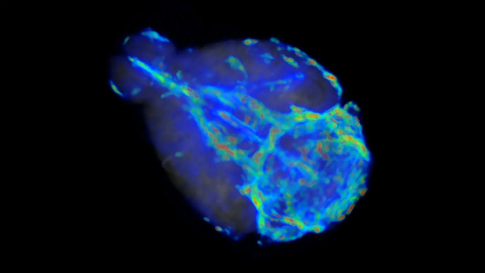
During the profound restorative stages of deep sleep, a crucial biological process unfolds within the intricate architecture of the brain: the efficient removal of metabolic byproducts. This cleansing operation, facilitated by a cerebrospinal fluid-like substance that circulates throughout the cerebral landscape, plays a vital role in mitigating the accumulation of waste materials linked to neurodegenerative conditions such as Alzheimer’s disease. This sophisticated waste management system, known as the glymphatic system, was first elucidated in 2012 by the groundbreaking work of Maiken Nedergaard, a distinguished neuroscientist and co-director of the University of Rochester’s Center for Translational Neuromedicine. While the fundamental existence of this system is established, the precise mechanisms governing its operation, particularly the speed at which fluid traverses brain tissues, have remained an area of intense scientific inquiry.
The challenge of precisely quantifying the movement of these fluids within the living brain presents a formidable hurdle for researchers. Traditional imaging techniques, while offering granular insights into localized regions, often fall short of providing a comprehensive, whole-brain perspective. Professor Douglas Kelley, a key figure in mechanical engineering at the University of Rochester, articulated this limitation, explaining that while direct microscopic observation of small brain areas can yield detailed data, it offers only a fragmented view of the overarching circulatory process. Magnetic Resonance Imaging (MRI), a powerful tool for generating three-dimensional brain maps, is ill-equipped to detect the exceedingly slow flow velocities characteristic of glymphatic circulation. This inherent constraint necessitated the development of novel methodologies capable of overcoming these observational barriers without inflicting damage on the delicate neural environment.
In response to this critical need, Professor Kelley, in collaboration with researchers from Brown University and the University of Copenhagen, harnessed the power of artificial intelligence to circumvent the limitations of conventional imaging. Their pioneering research, detailed in a recent publication in the esteemed journal Science Advances, introduces a novel physics-informed AI framework designed to calculate fluid flow speeds directly from MRI data. This innovative approach involved training sophisticated neural networks on a dataset of videos that meticulously documented the diffusion of tracer dyes through brain tissue over time. By meticulously analyzing the patterns of dye movement, the AI system gained the capacity to accurately estimate not only the velocity of the fluid but also the intricate permeability characteristics of the surrounding neural matrix.
The groundbreaking findings emerging from this AI-driven analysis have illuminated a fundamental duality in the brain’s waste removal strategy. The glymphatic system, it appears, operates through at least two distinct pathways, each characterized by dramatically divergent flow rates. These pathways are responsible for clearing a range of substances, including amyloid-beta proteins, which are strongly implicated in the pathogenesis of Alzheimer’s disease.
The more superficial regions of the brain, encompassing areas such as the perivascular spaces situated between the skull and the brain’s surface, exhibit a relatively swift fluid circulation, with the aqueous medium traversing at speeds measured in microns per second. However, as the fluid penetrates deeper into the intricate network of brain tissue, its velocity undergoes a significant deceleration. The research team discovered that this deep-tissue flow is approximately fifty times slower than its superficial counterpart, highlighting a distinct functional specialization within the glymphatic network.
Currently, the research endeavors are focused on establishing normative baselines for fluid dynamics within the brains of animal models, primarily mice. These baseline measurements are instrumental in refining and enhancing the accuracy of the developed AI tools. The ultimate objective is to leverage these refined tools to conduct comparative analyses of fluid circulation patterns between healthy and diseased brains, as well as to investigate age-related variations in this critical physiological process.
A significant long-term aspiration of this research is to translate these advanced methodologies to the study of the human brain. The ability to precisely measure fluid circulation within and around the human cerebrum holds immense potential for revolutionizing the diagnosis and understanding of neurological disorders and traumatic brain injuries. Professor Kelley expressed optimism regarding the clinical implications, stating that the ability to measure fluid flow in human brains would unlock more profound and exciting applications. The hope is to one day be able to ascertain whether individuals diagnosed with Alzheimer’s disease exhibit compromised cerebral circulation, or to implement early screening for such deficits to potentially preempt the onset of the condition. Furthermore, this technology could be employed to assess the impact of concussive injuries on brain fluid circulation, offering a crucial diagnostic tool. This recent study represents a significant stride toward achieving these ambitious clinical goals.
The scientific exploration underpinning this advancement has received crucial financial support from the NIH National Center for Complementary and Integrative Health and the NIH BRAIN Initiative, underscoring the national importance placed on understanding brain function and health. The collaborative nature of this research is evident in the diverse team of scientists involved. Professor Kelley’s key collaborators on this pivotal study include Juan Diego Toscano, a PhD student at Brown University; Yisen Guo, a computational scientist at the University of Rochester; Zhibo Wang, another PhD student from Brown University; Mohammad Vaezi, a PhD student at the University of Rochester; Yuki Mori, an Associate Professor at the University of Copenhagen; George Karniadakis, a Professor at Brown University; and Kimberly Boster, an Assistant Professor at the University of Rochester. This multidisciplinary effort highlights the complex and integrated approach required to unravel the intricate workings of the human brain.
About the Author












