
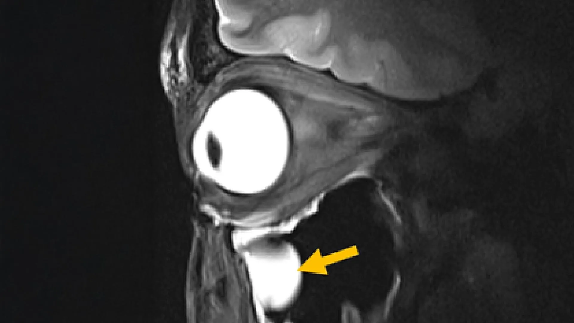
Magnetic Resonance Imaging (MRI), a cornerstone of modern medical diagnostics, has long grappled with the challenge of capturing exceptionally clear and detailed images of certain anatomically intricate or deeply situated bodily structures. The inherent limitations in current MRI hardware, particularly concerning the transmission and reception of radiofrequency (RF) signals, have historically presented obstacles to achieving optimal image quality in areas such as deep brain regions and the delicate tissues comprising the eye and its surrounding orbital structures. Addressing this persistent hurdle, a pioneering team, spearheaded by doctoral candidate Nandita Saha under the guidance of Professor Thoralf Niendorf at the Max Delbrück Center’s Experimental Ultrahigh Field Magnetic Resonance laboratory, has engineered a novel MRI antenna. This innovative device leverages advanced engineered materials to generate sharper images with reduced scanning times and boasts seamless integration into existing MRI systems, obviating the need for costly and disruptive equipment overhauls. The groundbreaking findings detailing this advancement have been formally presented in the esteemed journal Advanced Materials.
The collaborative effort behind this breakthrough united a diverse group of specialists, drawing expertise from the fields of MRI physics, clinical ophthalmology, and translational imaging. Contributions came from both the Max Delbrück Center and Rostock University Medical Center, with researchers in Rostock actively engaged in the crucial validation phase of the technology for its eventual clinical deployment. Professor Niendorf, the senior author of the publication, elaborated on the conceptual underpinnings of their innovation, stating, "By drawing upon principles from metamaterials, we successfully achieved a more efficient manipulation of radiofrequency fields, thereby demonstrating the direct impact advanced physics can have on refining medical imaging capabilities." He further emphasized the broad implications of their work, noting, "This research illuminates a viable path toward expediting MRI scans and enhancing their clarity, a development with the potential to significantly benefit patients across a multitude of clinical disciplines."
The fundamental operation of MRI scanners involves the emission of RF signals into the body while it is subjected to a potent magnetic field. The subsequent response of various tissues to these signals provides the data necessary for image reconstruction. Generally, a stronger signal translates to a more detailed and higher-resolution scan. However, conventional MRI antennas, also referred to as RF coils, frequently encounter difficulties in efficiently gathering adequate signal strength from tissues located deep within the body or situated in anatomically complex regions. This signal attenuation can compromise image quality and necessitate longer examination periods.
To surmount these limitations, the researchers strategically integrated metamaterials directly into the design of the MRI antenna. Metamaterials are a class of artificially engineered structures possessing electromagnetic properties that transcend those found in naturally occurring materials. In rigorous testing, the newly developed antenna exhibited a marked improvement in signal amplification from targeted tissues, a notable increase in spatial resolution, a perceptible enhancement in image sharpness, and a significant acceleration in the rate of data acquisition. A particularly compelling advantage of this innovation is its inherent compatibility with contemporary MRI hardware, thereby circumventing the substantial financial investment typically associated with acquiring entirely new infrastructure. The efficacy of this design was empirically validated through imaging the human eye and orbit in volunteer participants utilizing a 7.0 Tesla MRI scanner.
Professor Oliver Stachs, a co-author of the study affiliated with University Medicine Rostock, highlighted the profound relevance of this research for ophthalmic applications. He remarked, "Our investigation unequivocally demonstrates a clear utility for ophthalmological applications, as it paves the way for MRI of the eye with exceptional anatomical detail and high spatial resolution." Professor Stachs further articulated the transformative potential, stating, "This technology offers the prospect of opening an unprecedented window into the eye, allowing for the visualization of (patho)physiological processes that have historically remained largely inaccessible."
The overarching objective of the project, as articulated by Nandita Saha, was to "reimagine MRI hardware through the lens of modern antenna design physics." Saha further elaborated on the technology’s broader applicability, suggesting its adaptation for safeguarding sensitive bodily areas during MRI examinations by mitigating unwanted thermal effects around implanted medical devices. Additionally, the enhanced precision in directing RF energy could prove invaluable for MRI-guided cancer therapies, such as tumor hyperthermia or thermal tissue ablation, leading to more targeted and effective treatments.
MRI examinations, often characterized by their prolonged duration and potential for patient discomfort, can be further complicated by the need for repeat scans due to insufficient capture of critical anatomical details. The introduction of this new antenna, capable of producing superior images at an accelerated pace, promises to significantly reduce scan times while simultaneously instilling greater confidence in diagnostic accuracy for physicians. The antenna’s compact and lightweight construction also allows for customization tailored to specific anatomical regions, thereby contributing to enhanced patient comfort during imaging sessions. Professor Niendorf expressed optimism regarding the future scope of this technology, envisioning its adaptation for MRI systems operating across a spectrum of magnetic field strengths, both lower and higher than the 7.0 T used in initial testing. Furthermore, he anticipates its application in imaging organs beyond the eye, orbit, and brain, as well as in monitoring metabolic processes and tracking the pharmacokinetic behavior of pharmaceuticals within the body. The technology also holds promise for augmenting specialized MRI techniques that focus on imaging atomic nuclei other than hydrogen, such as sodium and fluorine, by yielding more robust signals and higher image fidelity. Dr. Ebba Beller, a co-author from Rostock University Medical Center, underscored the impact of hardware advancements, stating, "Innovations in imaging hardware possess the latent capacity to revolutionize diagnostic capabilities, and this study represents a pivotal stride towards the development of next-generation MRI technology."
The research consortium is currently embarking on the planning stages for more extensive clinical studies involving a multi-institutional collaboration. Concurrently, efforts are underway to refine the antenna’s design for the imaging of additional vital organs, including the heart and kidneys. The enduring scientific partnership between Professor Stachs and Professor Niendorf is set to continue, bolstered by reciprocal visiting scientist appointments, fostering ongoing interdisciplinary exchange and further innovation. This pioneering research was generously supported by the German Research Foundation (DFG) as a joint endeavor between the Max Delbrück Center and the Medical University Rostock.
About the Author












