
The intricate construction of the brain, a process of unparalleled biological complexity, involves a remarkable journey for its fundamental units: neurons. As these nascent nerve cells proliferate and differentiate, they embark on a migratory expedition, navigating a dense and increasingly crowded environment within the developing cerebral cortex. This passage is not a gentle stroll; it necessitates a forceful traversal through confined interstitial spaces, often squeezing between delicate cellular fibers and the tightly packed bodies of their burgeoning neighbors. This arduous physical undertaking, essential for populating the brain’s sophisticated communication network, has now been revealed to carry a profound and previously unrecognized consequence for the very genetic material of these migrating cells.
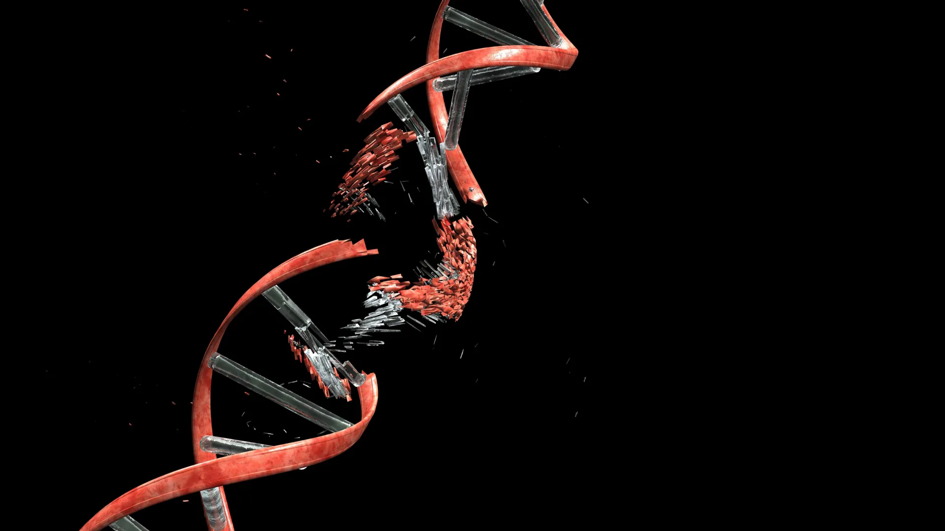
A groundbreaking investigation, recently detailed in the prestigious journal Nature, has illuminated an unexpected aspect of this developmental process. Scientists affiliated with Kyoto University’s Institute for Integrated Cell-Material Sciences (WPI-iCeMS), in collaboration with a consortium of esteemed research institutions, have uncovered compelling evidence that migrating neurons routinely sustain significant damage to their deoxyribonucleic acid (DNA). More specifically, these developing cells are observed to undergo double-strand breaks, a particularly severe form of genomic injury where both complementary strands of the DNA double helix are severed.
While double-strand breaks in DNA are widely recognized as potent instigators of genetic mutations, cellular dysfunction, and even programmed cell death (apoptosis), this new research posits a startling dichotomy. In the context of healthy brain cortex development, these breaks are not aberrations but rather integral, albeit temporary, components of the architectural assembly process. Crucially, the study demonstrates that in the pristine environment of a developing brain, these damaging events are subject to rapid and highly efficient repair mechanisms. This swift restoration of genomic integrity ensures that the transient structural alterations do not precipitate lasting cellular pathologies or functional impairments.
Professor Mineko Kengaku, a leading figure at WPI-iCeMS and the principal investigator of this pivotal study, articulated the study’s core insight: "The developing brain appears to have evolved a remarkable capacity to both tolerate and proficiently mend the DNA damage encountered by migrating neurons." She further elaborated on the significance of this finding, stating, "However, a deeper comprehension of the boundaries of this tolerance, and more importantly, the downstream effects when this repair process is compromised or incomplete, brings us measurably closer to deciphering the underlying mechanisms of a spectrum of neurological conditions."
To meticulously dissect the genesis of this neuronal DNA damage, the research team ingeniously devised experimental conditions that mirrored the physical tribulations faced by developing neurons in vivo. They engineered sophisticated microfluidic channels, meticulously calibrated in their dimensions to replicate the constricted passages characteristic of growing brain tissue. Neurons were then guided through these artificial conduits, simulating their natural migratory path.
Employing advanced fluorescent tagging techniques, the researchers were able to visualize and track the emergence of double-strand DNA breaks in real-time as neurons traversed these confined microchannels. The observations were striking: upon successfully navigating these tight spaces and emerging into more open environments, the incidence of DNA damage began to recede. The study quantifies this recovery, noting that the majority of these breaks were successfully repaired within a 24-hour period, allowing the neurons to resume their normal functional trajectories without apparent detriment.
The investigation delved deeper to pinpoint the molecular culprit behind this transient genomic insult. The researchers identified a critical enzyme, Topoisomerase IIβ (TopoIIβ), as the primary agent involved. This enzyme typically plays a vital role in managing the inherent mechanical stresses within DNA molecules that arise from routine cellular activities. Under normal physiological circumstances, TopoIIβ functions by transiently cleaving DNA strands, thereby alleviating torsional strain and preventing entanglement, before meticulously rejoining the severed ends. This action can be conceptually likened to temporarily cutting a twisted cable to release tension and then expertly splicing it back together. However, the study reveals that when neurons are subjected to significant mechanical forces while navigating extremely narrow passages, TopoIIβ can become inadvertently trapped in its catalytic cycle, leaving the DNA strands broken and unrepaired. In such instances, the cell is compelled to rely on an alternative repair pathway, known as non-homologous end joining (NHEJ), to bridge the fractured DNA termini.
A crucial aspect of the research focused on elucidating why neurons exhibit such resilience to DNA damage, a characteristic not universally shared by other cell types. The study drew a salient contrast between the DNA damage observed in migrating neurons and that occurring in certain types of cancer cells when subjected to similar microchannel confinement. In neoplastic cells, DNA damage often manifests as more widespread and erratic, frequently disrupting essential cellular functions or even triggering cell death pathways.
Conversely, the DNA breaks in neurons were found to be predominantly localized to specific regions of the genome that are not actively engaged in critical gene expression. This strategic sparing of essential genetic machinery allows neurons to maintain their core functionality despite the temporary, localized genomic disruptions. By avoiding damage to vital genes, the cells retain their capacity to develop and integrate into neural circuits.
The implications of impaired DNA repair mechanisms were further explored through a series of experiments involving genetically engineered mice. These mice were specifically designed to have newly formed cerebellar neurons that were deficient in Ligase 4, an enzyme indispensable for the proper execution of the NHEJ DNA repair pathway. Intriguingly, these mice exhibited normal development and showed no overt signs of abnormality during their early life stages. However, as they matured into adulthood, a subtle yet progressively worsening deficit in balance and coordination became apparent. These motor impairments bear a striking resemblance to the clinical manifestations observed in certain human genetic disorders characterized by genome instability, particularly those affecting the cerebellum, a brain region critical for motor control and coordination.
These findings collectively suggest that the intricate processes of DNA breakage and subsequent repair may exert a far more significant influence on fundamental brain biology than had been previously appreciated. The researchers now aim to investigate whether these early-life genomic alterations contribute to the observed heterogeneity among individual neurons and, furthermore, whether they play a role in the pathogenesis of neurodevelopmental disorders, such as autism spectrum disorder or attention-deficit/hyperactivity disorder, and neurodegenerative diseases that manifest later in life, such as Alzheimer’s or Parkinson’s disease.
Professor Kengaku underscored the transformative nature of these discoveries, stating, "This research fundamentally shifts our perspective on the neuronal genome. While all neurons ostensibly originate from the same genetic blueprint, the dynamic interplay of DNA damage and repair, particularly during the critical migratory phase, can introduce subtle yet meaningful genetic variations between individual neurons. It is conceivable that a portion of this developmental history, etched into the genome itself, may ultimately influence neuronal identity and function throughout an organism’s lifespan."
This extensive collaborative effort involved contributions from researchers at the University of Tokyo, Osaka University, the National University of Singapore, and the Tokyo Metropolitan Institute of Medical Science, in addition to the lead institution, Kyoto University.
About the Author












