
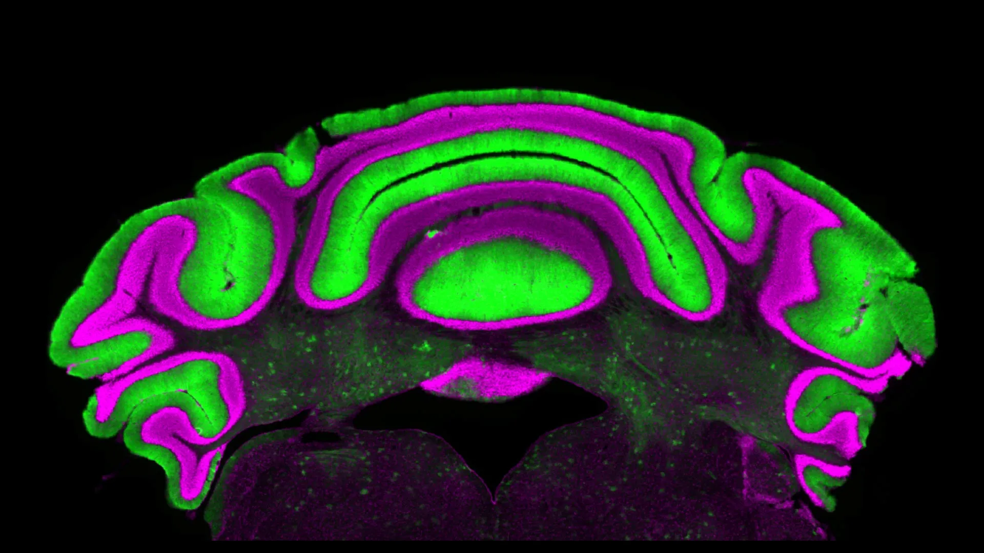
A groundbreaking investigation conducted by neuroscientists at the Fralin Biomedical Research Institute at Virginia Tech is challenging established paradigms in the scientific exploration of chronic neurological conditions, including dystonia, ataxia, and tremors. These debilitating disorders, which manifest as involuntary muscle contractions, postural abnormalities, and persistent shaking, are intrinsically linked to disruptions within the cerebellum, a crucial brain region responsible for motor coordination and fine-tuning movements. For a considerable period, the scientific community has largely operated under the assumption that the activity patterns of one specific neuronal type within the cerebellum, namely Purkinje cells, directly and reliably reflect the functional state of another, the deep cerebellar nuclei cells. This long-held belief has underpinned many research methodologies and therapeutic strategies.
Purkinje cells, characterized by their extensive dendritic trees and strategic location in the outer granular layer of the cerebellum, have been a focal point for researchers due to their relative accessibility for study. In contrast, deep cerebellar nuclei cells are situated deeper within the brain structure, posing greater challenges for direct physiological measurement. Consequently, the activity of Purkinje cells has often been interpreted as a readily available proxy or biomarker for the activity of these more deeply embedded neuronal populations. This indirect observational approach has facilitated extensive research into the mechanisms of cerebellar function and dysfunction. The prevailing scientific consensus posited a clear inhibitory relationship: heightened activity in Purkinje cells would predictably lead to suppressed activity in the deep cerebellar nuclei, and conversely, diminished Purkinje cell firing would result in increased activity in the nuclei. This presumed linear correlation served as a foundational principle for many experimental designs and therapeutic interventions aimed at modulating cerebellar output.
However, the recent study, spearheaded by Assistant Professor Meike van der Heijden and published in the esteemed Journal of Physiology, presents a compelling body of evidence that calls into question this fundamental assumption. The research team’s meticulous analysis of electrophysiological recordings, derived from preclinical models exhibiting cerebellar pathologies, yielded results that were both surprising and significant. Contrary to expectations, the study found no discernible or consistent correlation between the firing rates of Purkinje cells and deep cerebellar nuclei cells. This finding directly challenges the long-standing hypothesis that monitoring Purkinje cell activity can accurately predict or represent the state of the deep nuclei.
"We observed that there isn’t a clear, linear relationship between the activity of Purkinje cells and that of the deep nuclei cells," explained Van der Heijden, who also holds an appointment within Virginia Tech’s School of Neuroscience. "This means there is very limited predictive power in monitoring one to understand what is happening in the other." This revelation suggests that the intricate circuitry and signaling within the cerebellum may be far more complex and nuanced than previously understood, with distinct neuronal populations exhibiting independent or less directly coupled operational dynamics.
The implications of these findings are profound and far-reaching, potentially necessitating a substantial revision of how movement disorders are investigated and treated. Alyssa Lyon, a doctoral candidate in Virginia Tech’s Translational Biology, Medicine, and Health Graduate Program and the primary author of the study, elaborated on the significance. "Purkinje and cerebellar deep nuclei cell activity is disrupted in a disease state, and a better understanding of the relationship between these neuron types will ultimately help optimize treatments for diseases such as dystonia, ataxia, and tremor," she stated. The current reliance on Purkinje cell activity as a surrogate indicator for deep nuclei function may have led to incomplete or even misdirected research efforts, potentially hindering the development of more effective therapies.
The study’s methodology involved a comprehensive examination of a large dataset of electrophysiological recordings from animal models designed to mimic the conditions seen in human cerebellar movement disorders. By analyzing these recordings, the researchers were able to assess the simultaneous activity of both Purkinje cells and deep cerebellar nuclei cells under various experimental conditions. The consistent absence of a significant correlation across these diverse datasets strongly suggests that the presumed direct inhibitory linkage does not translate into a predictable functional relationship in practice, particularly in the context of disease states.
Van der Heijden emphasized the critical need for a paradigm shift in research approaches. "If you want to understand how the cerebellum is functioning in a disease state, you must look at the deep nuclei neurons, not just the Purkinje cells," she asserted. This recommendation underscores the importance of direct measurement and investigation of the deep cerebellar nuclei, despite the inherent technical challenges. Future research endeavors will likely need to incorporate more sophisticated techniques for accessing and recording from these deeper neuronal populations to gain a more accurate understanding of their roles in health and disease.
Furthermore, the study serves as a crucial cautionary note for the development of therapeutic strategies. Many existing or proposed treatments for cerebellar movement disorders aim to modulate Purkinje cell activity, with the implicit expectation that this modulation will lead to the desired downstream effects in the deep cerebellar nuclei. The new findings suggest that such interventions might not achieve their intended outcomes if the underlying assumption of a direct, predictable link is flawed. "This is a cautionary tale for understanding cerebellar activity in disease, but also for treating these challenging diseases," Van der Heijden added. "We need to be very careful in making assumptions, and to actually do experiments to test our hypotheses."
The discovery highlights the inherent complexity of neural circuits and the potential pitfalls of oversimplification in scientific inquiry. While Purkinje cells play an undeniably vital role in cerebellar function, their activity alone may not be a sufficient indicator of the overall functional status of the cerebellar output pathways. The deep cerebellar nuclei are the primary output structures of the cerebellum, projecting to numerous brain regions involved in motor control, cognition, and emotion. Therefore, understanding the precise dynamics of these nuclei is paramount for deciphering the origins of movement disorders and for devising targeted and effective interventions.
This research opens new avenues for scientific exploration, encouraging a more holistic and detailed examination of cerebellar circuitry. It underscores the imperative for empirical validation of long-held hypotheses and advocates for the adoption of methodologies that provide a more direct and comprehensive view of neuronal function. The implications for patients suffering from dystonia, ataxia, and tremor are substantial, as a refined understanding of cerebellar mechanisms could pave the way for novel therapeutic approaches that are more precisely tailored to the underlying pathophysiology of these conditions. The scientific community is now tasked with re-evaluating established models and embracing more intricate investigative techniques to unlock the full potential of cerebellar research and improve the lives of those affected by movement disorders. The unexpected findings serve as a potent reminder that scientific progress often hinges on the willingness to challenge existing dogma and pursue new, sometimes unconventional, lines of inquiry.
About the Author












