
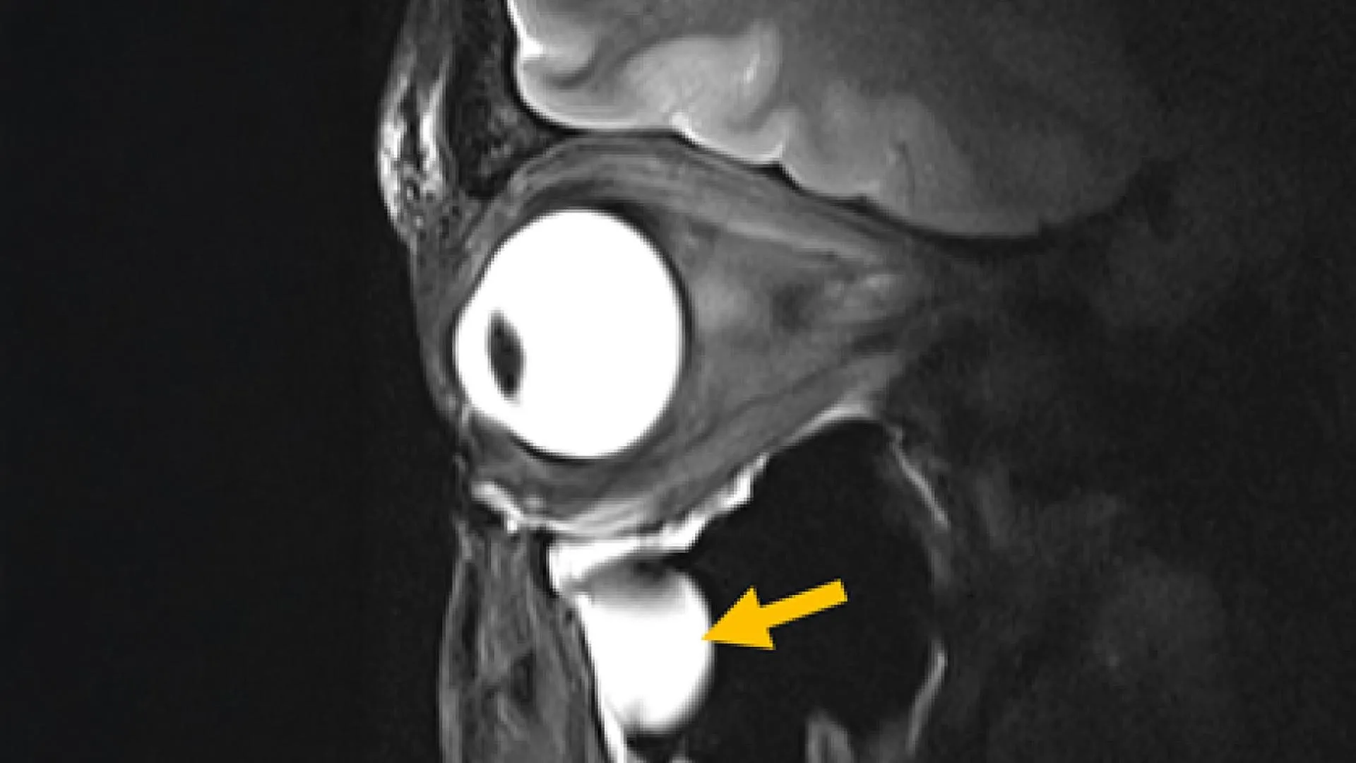
Magnetic Resonance Imaging (MRI), a cornerstone of modern medical diagnostics, has long been celebrated for its non-invasive ability to visualize internal bodily structures. However, the inherent physics of radiofrequency signal transmission and reception has historically presented significant challenges when attempting to generate high-fidelity images of certain anatomical regions. Specifically, the intricate architecture of deep brain structures and the exquisitely sensitive tissues within and surrounding the eye have proven particularly recalcitrant to detailed visualization, often yielding images with compromised resolution or requiring extended scanning times. Addressing this persistent limitation, a pioneering development by a dedicated research consortium promises to redefine the boundaries of MRI’s diagnostic power.
At the vanguard of this advancement is a novel MRI antenna, meticulously engineered by a team spearheaded by Nandita Saha, a doctoral candidate within Professor Thoralf Niendorf’s renowned Experimental Ultrahigh Field Magnetic Resonance laboratory at the Max Delbrück Center. This innovative antenna leverages the unique properties of advanced, specially designed materials to achieve a remarkable leap in image quality and acquisition speed. Crucially, its design is intrinsically compatible with existing MRI systems, obviating the need for substantial and costly overhauls of current clinical infrastructure. The groundbreaking findings detailing this achievement have been formally disseminated through publication in the esteemed journal Advanced Materials.
The collaborative spirit driving this innovation is a testament to interdisciplinary synergy, uniting leading minds from the fields of MRI physics, clinical ophthalmology, and translational imaging. Researchers from both the Max Delbrück Center and the Rostock University Medical Center have joined forces, with their counterparts in Rostock actively engaged in the rigorous validation processes essential for the technology’s eventual translation into widespread clinical practice.
Professor Thoralf Niendorf, the senior author of the published research, articulated the fundamental principle underpinning their success: "By drawing upon concepts derived from metamaterials, we have successfully devised methods to channel radiofrequency fields with substantially enhanced efficiency, thereby demonstrating a direct pathway through which advanced physics can profoundly improve the quality of medical imaging." He further elaborated on the broader implications, stating, "This work illuminates a promising trajectory toward MRI scans that are not only faster but also significantly clearer, a development poised to offer tangible benefits to patients across a multitude of clinical disciplines."
The intricate process of MRI image generation relies on the precise interplay between powerful magnetic fields and carefully modulated radiofrequency (RF) signals. The body’s tissues respond to these stimuli by emitting signals that the MRI scanner meticulously collects, piecing together a detailed anatomical representation. Intuitively, a stronger and more refined signal translates directly into sharper, more informative images. However, traditional RF coils, the components responsible for transmitting and receiving these critical signals, often struggle to capture sufficient signal strength from tissues situated deep within the body or residing in regions characterized by complex anatomical configurations. This inherent difficulty can lead to a noticeable degradation in image quality and necessitate prolonged scanning periods, impacting both diagnostic accuracy and patient comfort.
The breakthrough achieved by Saha and her colleagues lies in the ingenious integration of metamaterials directly into the fabric of the MRI antenna. Metamaterials are not naturally occurring substances; rather, they are sophisticated artificial structures meticulously designed to exhibit electromagnetic properties that deviate markedly from those found in conventional materials. In rigorous experimental testing, the newly developed antenna demonstrably amplified signals emanating from targeted tissues, concurrently enhancing spatial resolution, refining image sharpness to an unprecedented degree, and accelerating the rate at which data could be acquired.
A particularly significant advantage of this novel antenna is its seamless integration capability with existing MRI hardware. This compatibility effectively removes the substantial financial barrier associated with the acquisition of entirely new, state-of-the-art imaging machines, making advanced imaging accessible to a broader range of healthcare institutions. The research team put their innovation to the test by successfully imaging the intricate structures of the human eye and its surrounding orbit in volunteer participants, utilizing a high-field strength 7.0 Tesla MRI scanner.
Professor Oliver Stachs, a key co-author from the University Medicine Rostock, emphasized the profound implications for ophthalmology: "Our research provides unequivocal evidence of its substantial relevance for ophthalmic applications, offering the capacity to facilitate MRI scans of the eye with exceptional anatomical detail and high spatial resolution." He further highlighted the transformative potential, envisioning it as "an opening into the eye, providing unprecedented visibility into (patho)physiological processes that have, until now, remained largely beyond our reach."
The genesis of this project, as described by Saha, was rooted in a fundamental re-evaluation of MRI hardware design principles: "Our overarching objective was to fundamentally rethink MRI hardware through the lens of modern antenna design physics."
Beyond its immediate application in ocular imaging, Saha indicated that the technology’s adaptability extends to several other critical areas within MRI. It holds promise for enhancing patient safety during MRI examinations by mitigating the potential for unwanted tissue heating in proximity to sensitive medical implants. Furthermore, it could significantly improve the precision and efficacy of MRI-guided cancer treatments by enabling more targeted delivery of RF energy for procedures such as tumor hyperthermia or thermal tissue ablation.
The often lengthy and potentially uncomfortable nature of MRI examinations, particularly when repeated scans are required due to insufficient detail in initial images, represents a significant challenge in clinical workflow. By delivering superior image clarity at an accelerated pace, the new antenna offers a compelling solution, promising to shorten examination durations while simultaneously bolstering physicians’ confidence in their diagnostic conclusions. The antenna’s compact and lightweight design also facilitates customization for different anatomical regions, potentially enhancing patient comfort during the imaging process.
Professor Niendorf further projected the technology’s future trajectory, suggesting its eventual adaptation for MRI systems operating across a wider spectrum of magnetic field strengths, both lower and higher than the 7.0 T used in initial testing. The potential applications could extend to imaging organs beyond the eye, orbit, and brain, and even be employed for monitoring metabolic processes and tracking the pharmacokinetics of drug distribution within the body. Additionally, this innovation could significantly advance specialized MRI techniques that focus on imaging atoms other than hydrogen, such as sodium and fluorine, by generating stronger signals and yielding higher quality diagnostic images.
Dr. Ebba Beller, another co-author from Rostock University Medical Center, underscored the broader significance of such hardware advancements: "Innovations in imaging hardware possess the profound capability to revolutionize diagnostic methodologies, and this study represents a pivotal step toward the realization of next-generation MRI technology."
Looking ahead, the research team is actively embarking on more extensive clinical studies, enlisting the participation of multiple medical institutions. Concurrently, they are refining the antenna design for its application to other vital organs, including the heart and kidneys. The established and fruitful collaboration between Professor Stachs and Professor Niendorf is set to continue, further strengthened by reciprocal visiting scientist appointments that foster ongoing knowledge exchange and collaborative research endeavors. This pioneering work was made possible through funding from the German Research Foundation (DFG), representing a significant joint effort between the Max Delbrück Center and the Medical University Rostock.
About the Author












