Retinal Circuitry Revealed: Yale Researchers Uncover Hidden Interconnectivity Enhancing Visual Perception
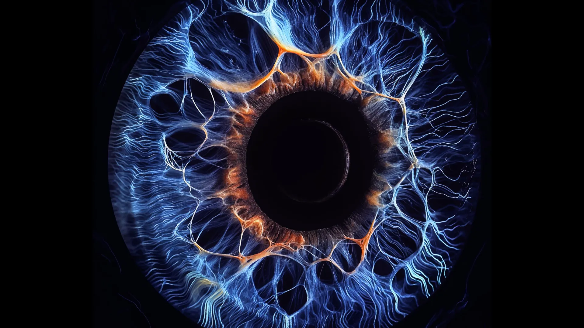
A groundbreaking investigation originating from Yale School of Medicine (YSM) has unveiled a sophisticated, previously unrecognized communication infrastructure within the eye, fundamentally altering our comprehension of how visual information is processed. This discovery challenges long-held assumptions about the segregation of visual pathways and offers compelling explanations for our capacity to discern subtle visual cues and navigate in low-light environments. The findings suggest a more dynamic and integrated system than previously envisioned, where distinct sensory streams are not as isolated as once believed.
The human visual apparatus is renowned for its remarkable speed and efficiency in dissecting the myriad elements of a scene, encompassing attributes such as hue, luminosity variations, movement, and geometric form. This multifaceted analytical capability, often referred to as parallel visual processing, empowers the brain to interpret intricate imagery with near-instantaneous speed. The prevailing scientific consensus had posited that this parallel processing involved separate, largely independent conduits, or pathways, through which visual signals traversed the retina en route to the brain. However, the YSM study, meticulously detailed in the esteemed scientific journal Neuron, presents compelling evidence of pervasive and intricate links between these presumed independent channels, facilitated by concealed electrical connections. The research team posits that this cooperative intercommunication serves to amplify feeble visual signals, thereby bolstering their integrity before they advance further into the visual processing hierarchy.
Dr. Yao Xue, a postdoctoral fellow in YSM’s Department of Ophthalmology and Visual Science and the lead author of the study, articulated the core finding: "We discovered that while distinct channels are indeed capable of conveying their specific feature information, they are simultaneously interconnected through an underlying electrical circuitry." This suggests a fundamental shift in understanding, moving from a model of siloed processing to one of synergistic collaboration.
The intricate cascade of vision commences with photoreceptor cells, the rods and cones situated within the retina, which are responsible for transducing light into neural signals. These initial signals are then relayed to a class of neurons known as bipolar cells. At this juncture, the raw visual information undergoes a crucial initial sorting process, bifurcating into more than a dozen distinct parallel channels. Each of these channels is specialized to process specific aspects of the visual input, such as those relevant to diurnal or nocturnal vision, color differentiation, contrast sensitivity, and shape recognition.
Through rigorous microscopic examination of the synapses – the minute junctions where bipolar cells transmit signals to one another – the researchers encountered a profound and unanticipated revelation. Far from maintaining their presumed isolation, these ostensibly separate informational channels were found to be actively exchanging data. Neuronal communication primarily occurs via two principal mechanisms: chemical synapses, which employ neurotransmitters as chemical messengers, and electrical synapses, also termed gap junctions, which facilitate the direct passage of electrical currents between cells. Until this study, bipolar cells were generally understood to rely predominantly on chemical signaling for their interactions.
However, the YSM investigation, conducted on both rodent and human retinal tissues, unequivocally demonstrated the widespread presence of electrical synapses bridging the majority of these segregated information pathways. A pivotal experimental maneuver involved the targeted electrical stimulation of a single bipolar cell. The resulting neural response was observed to propagate far beyond the confines of its originating pathway, manifesting as broad, diffuse patterns of activity rather than localized neurotransmitter release. This observation served as a striking indicator of extensive communication and information sharing among different types of bipolar cells.
The principal investigator of the study, Dr. Z. Jimmy Zhou, the Marvin L. Sears Professor of Ophthalmology and Visual Science, elaborated on this observation: "When we stimulated one bipolar cell, a multitude of bipolar cells subsequently released neurotransmitters." This finding points to a collective response, rather than an isolated event.
Furthermore, the research team identified a specific subtype of bipolar cell, designated as BC6, that appeared to assume a central role in orchestrating this intricate network. Signals originating from BC6 exhibited a tendency to propagate across multiple visual pathways in a structured, hierarchical fashion, suggesting a leadership or coordinating function within the circuit. Dr. Zhou commented on this aspect: "Previously, it was assumed that the different types of bipolar cells operated in a largely autonomous manner. However, we have identified a key driver among these cell types that orchestrates this network with a defined hierarchy."
The synergy between specialized processing channels and this newly discovered electrical communication system, according to the scientists, endows the retina with an optimal combination of advantages. The specialized channels can meticulously focus on discerning particular visual features, while their interconnections enable the efficient pooling of information, particularly when dealing with exceptionally faint signals. Dr. Seunghoon Lee, a research scientist in the Department of Ophthalmology and Visual Science and co-corresponding author, explained the significance: "If a signal is already very weak and is divided among several channels, there is very little information left for each individual channel to process. This integration is especially beneficial for detecting low-contrast signals or stimuli originating from very small objects." He further added, "And importantly, the cells are not cooperating in a random fashion. There is a commander within them – BC6 – that guides them in relaying signals to the downstream target."
The sophisticated mapping of these communication networks was achieved through the integration of several advanced experimental methodologies. The researchers employed cutting-edge imaging techniques to meticulously monitor the release and reception of neurotransmitters by bipolar cells. Concurrently, they subjected individual cells to stimulation and recorded the resultant responses in neighboring cells. The study of bipolar cells has historically presented significant technical challenges due to their deep-seated location within the retinal layers. Prior experimental approaches often necessitated slicing the retina to access these cells, a procedure that could inadvertently disrupt the very neural circuitry under investigation.
In a significant methodological advancement, the Yale team successfully implemented a dual patch clamp technique on fully intact mouse retinas. Utilizing precisely positioned electrodes, they were able to stimulate specific types of bipolar cells while simultaneously recording the electrical responses of adjacent cells. Dr. Zhou highlighted the technical prowess involved: "No other laboratory in the world has been able to systematically perform these types of recordings. It represents a tour de force of Yao Xue’s doctoral thesis work, combining an innovative approach with exceptional electrophysiological skill." The team subsequently replicated these experiments using intact human retinas, obtained through the Legacy Tissue Donation Program of the Department of Pathology. According to the researchers, these represent the inaugural experiments of their kind ever conducted on an intact human retina.
The implications of this discovery extend beyond the realm of visual science. Given that the retina is an extension of the central nervous system, the researchers suggest that these findings could offer novel insights into the operational principles of other neural networks within the brain. Moreover, this enhanced understanding of retinal circuitry may significantly contribute to advancements in the diagnosis and treatment of diseases that compromise retinal health, such as age-related macular degeneration, glaucoma, and congenital night blindness. The study also serves as a powerful testament to the value of curiosity-driven scientific inquiry. Rather than pursuing a predetermined hypothesis, the experimental design allowed for the serendipitous uncovering of a fundamental, previously unknown mechanism that reshapes our understanding of visual processing. Dr. Lee concluded by emphasizing this point: "Our experiments did not commence with a specific hypothesis but rather revealed a fundamental processing mechanism within the visual system. It is an important reminder of how essential curiosity-driven research is to the process of discovery."

Related News