
The specific condition targeted by this advanced AI is acromegaly, an infrequently encountered disorder that typically emerges during middle age. Its underlying cause is the overproduction of growth hormone, a physiological anomaly that results in the gradual enlargement of extremities such as hands and feet. Acromegaly can also precipitate alterations in facial structure and promote abnormal growth patterns in skeletal tissues and internal organs. The insidious and protracted nature of its development, often spanning many years, presents a considerable challenge to its early identification.
Without timely and effective intervention, acromegaly is associated with a substantial increase in the risk of severe health complications and is estimated to reduce an individual’s life expectancy by approximately a decade. Dr. Hidenori Fukuoka, an endocrinologist at Kobe University and a lead researcher on the project, highlighted the diagnostic quandary, stating, "Because the condition progresses so slowly, and because it is a rare disease, it is not uncommon to take up to a decade for it to be diagnosed." He further elaborated on the historical context of technological adoption, noting, "With the progress of AI tools, there have been attempts to use photographs for early detection, but they have not been adopted in clinical practice." This statement underscores the persistent challenge of translating AI advancements into tangible clinical utility.
The genesis of this privacy-centric AI approach stemmed from a comprehensive review of existing artificial intelligence methodologies employed in medical diagnostics. The research team observed a prevailing trend wherein many AI systems relied heavily on facial photographs for disease identification. However, this reliance on facial data inherently introduces significant privacy concerns for patients, potentially hindering widespread adoption and patient willingness to participate. In direct response to this critical limitation, the scientific team deliberately charted a different course, opting for an alternative imaging strategy.
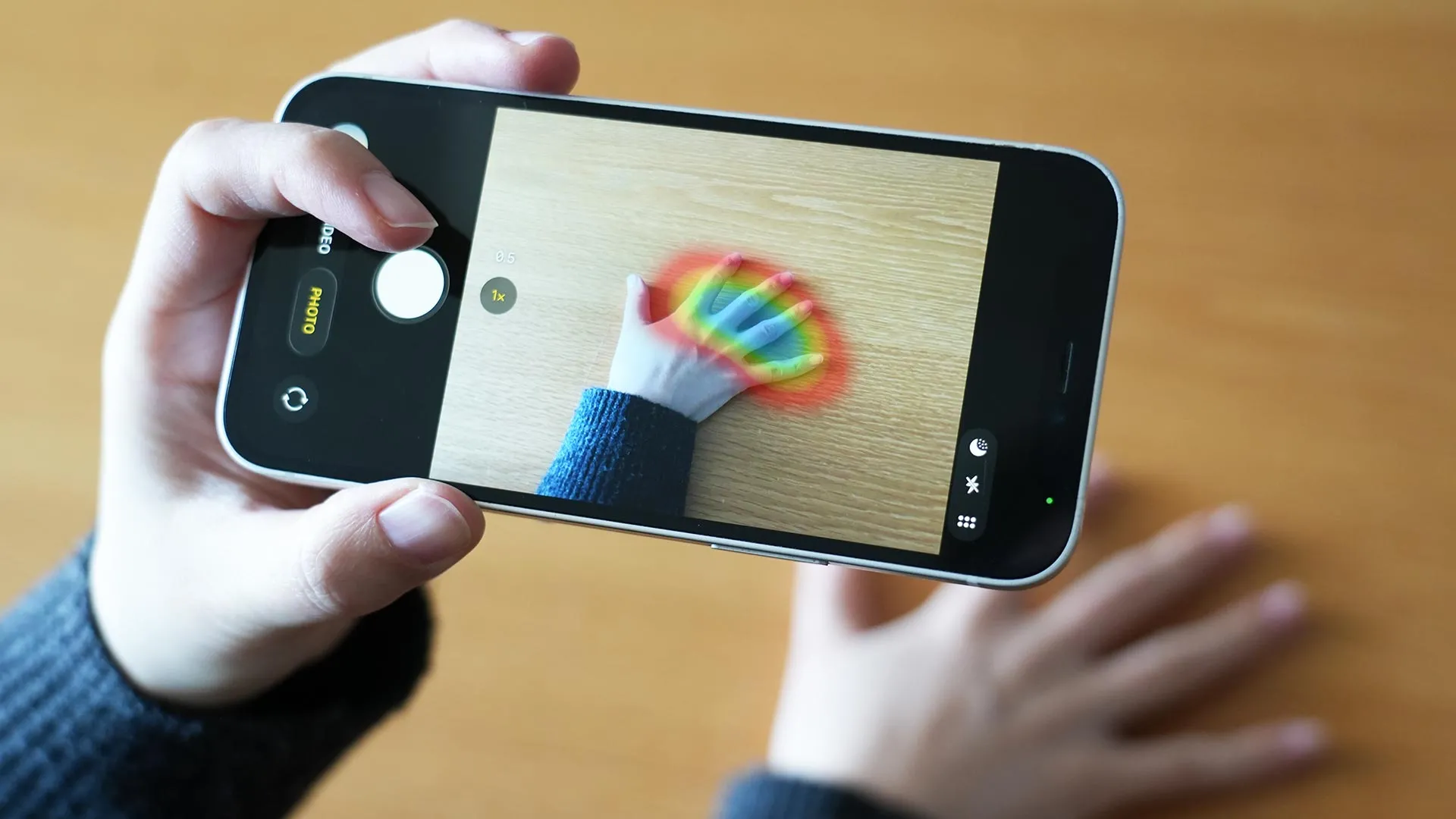
Yuka Ohmachi, a graduate student at Kobe University and a key contributor to the research, explained the rationale behind this strategic shift: "Trying to address this concern, we decided to focus on the hands, a body part we routinely examine alongside the face in clinical practice for diagnostic purposes, particularly because acromegaly often manifests changes in the hands." This decision was rooted in the understanding that hands, like the face, are frequently observed by clinicians for diagnostic clues, and importantly, that acromegaly exhibits characteristic manifestations in the hands.
To further bolster privacy protections and encourage broader participation, the researchers implemented a stringent imaging protocol. Their methodology was deliberately confined to capturing images of the dorsal aspect of the hand and a clenched fist. Crucially, they made a conscious decision to eschew photographs of the palm. This exclusion was driven by the highly individualized nature of palm lines, which could potentially serve as unique identifiers and compromise patient anonymity. This meticulously designed protocol proved instrumental in facilitating the recruitment of a substantial cohort of participants. Ultimately, the study amassed over 11,000 images contributed by 725 patients from 15 distinct medical institutions across Japan. This extensive dataset was subsequently utilized for the rigorous training and validation of the AI model.
In a remarkable demonstration of its efficacy, the research team’s AI model achieved exceptionally high levels of sensitivity and specificity in its ability to diagnose acromegaly from the captured hand images. These findings were formally presented in the prestigious Journal of Clinical Endocrinology & Metabolism. The performance metrics revealed that the AI system not only achieved superior accuracy but, in direct comparative evaluations, surpassed the diagnostic capabilities of experienced endocrinologists who were tasked with assessing the same set of photographs.
Ohmachi expressed her astonishment at the system’s capabilities, remarking, "Frankly, I was surprised that the diagnostic accuracy reached such a high level using only photographs of the back of the hand and the clenched fist. What struck me as particularly significant was achieving this level of performance without facial features, which makes this approach a great deal more practical for disease screening." This sentiment underscores the transformative potential of an AI diagnostic tool that can operate effectively without recourse to sensitive personal identifiers like facial data.
Looking beyond acromegaly, the researchers harbor ambitious aspirations to adapt and extend their AI system’s capabilities to encompass the detection of a wider spectrum of medical conditions that manifest observable changes in the hands. Potential future targets for investigation include conditions such as rheumatoid arthritis, anemia, and finger clubbing, all of which have distinct physical indicators in the hands. Ohmachi articulated this forward-looking vision, stating, "This result could be the entry point for expanding the potential of medical AI." This suggests a paradigm shift in how AI can be integrated into broader medical screening and diagnostic workflows.
Within the intricate landscape of actual clinical practice, physicians invariably integrate a multitude of diagnostic inputs beyond mere visual examination of hand photographs. A patient’s comprehensive medical history, the results of laboratory tests, and thorough physical examinations all play pivotal roles in forming a diagnostic conclusion. The Kobe University researchers position their AI tool not as a replacement for human medical expertise, but rather as a sophisticated adjunct designed to support and augment physician decision-making. In their published study, they aptly characterize the technology as a means to "complement clinical expertise, reduce diagnostic oversight and enable earlier intervention."
Lead investigator Dr. Fukuoka emphasized the long-term vision for the technology’s integration into healthcare infrastructure. "We believe that, by further developing this technology, it could lead to creating a medical infrastructure during comprehensive health check-ups to connect suspected cases of hand-related disorders to specialists," he stated. He further elaborated on its potential impact in underserved areas: "Furthermore, it could support non-specialist physicians in regional healthcare settings, thus contributing to a reduction of healthcare disparities there." This highlights the potential for the AI to democratize access to specialized medical insights, particularly in geographically remote or medically underserved regions. The research initiative was made possible through the generous financial support of the Hyogo Foundation for Science Technology. The collaborative nature of this groundbreaking project also involved significant contributions from a consortium of esteemed institutions, including Fukuoka University, Hyogo Medical University, Nagoya University, Hiroshima University, Toranomon Hospital, Nippon Medical School, Kagoshima University, Tottori University, Yamagata University, Okayama University, Hyogo Prefectural Kakogawa Medical Center, Hokkaido University, International University of Health and Welfare, Moriyama Memorial Hospital, and Konan Women’s University. This interdisciplinary collaboration underscores the complex and multifaceted nature of developing and validating advanced medical AI systems.
About the Author


