
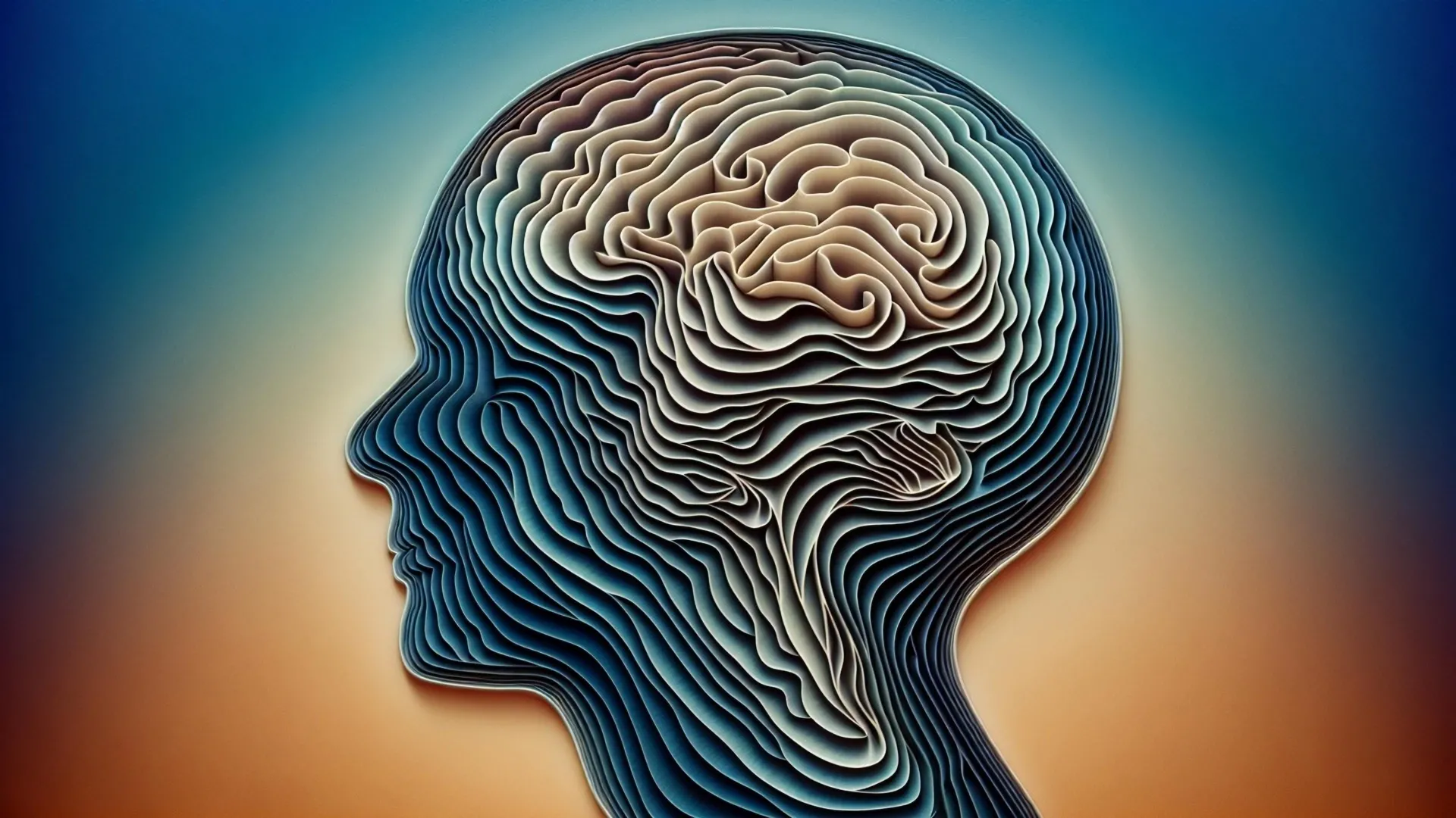
A groundbreaking investigation conducted by scientists at the Mark and Mary Stevens Neuroimaging and Informatics Institute (Stevens INI), affiliated with the Keck School of Medicine of USC, has illuminated a previously unrecognized architectural principle within a crucial area of the brain responsible for learning and memory. Their findings, meticulously detailed in the scientific journal Nature Communications, reveal that the CA1 sector of the hippocampus in mice is not a homogenous entity but rather comprises four distinct strata, each characterized by specialized cell populations. The hippocampus, a brain structure fundamental to memory formation, spatial orientation, and emotional processing, has now offered a deeper understanding of how neural information is transmitted through its intricate circuitry. This discovery also provides significant clues regarding the differential susceptibility of certain neuronal types to neurodegenerative conditions such as Alzheimer’s disease and neurological disorders like epilepsy.
For a considerable time, the scientific community has posited that disparate segments within the CA1 region of the hippocampus might be dedicated to distinct facets of learning and memory; however, the precise cellular arrangement underpinning this functional segregation remained elusive. The senior author of the study, Dr. Michael S. Bienkowski, an assistant professor of physiology and neuroscience and biomedical engineering at Stevens INI, articulated that their research has definitively demonstrated that CA1 neurons are organized into four slender, contiguous bands, with each band representing a unique neuronal subtype identifiable by its specific molecular fingerprint. He further elaborated that these layered structures are not static but exhibit subtle variations in their thickness and position along the longitudinal axis of the hippocampus. This dynamic, shifting pattern implies that each localized area of CA1 possesses a unique ensemble of neuron types, thereby offering a compelling explanation for why different hippocampal subregions are associated with diverse behavioral outputs. Moreover, this insight could potentially elucidate the differential vulnerability of specific CA1 neurons observed in conditions like Alzheimer’s disease and epilepsy; if a pathological process targets the cellular constituents of a particular layer, the resultant impact would invariably depend on the prominence of that layer in a given hippocampal locale.
The team leveraged an advanced RNA imaging technique known as RNAscope, coupled with high-resolution microscopy, to meticulously examine this intricate cellular architecture. This sophisticated methodology enabled the researchers to visualize gene expression at the single-molecule level within the CA1 tissue of mice, thereby facilitating the identification of individual neuron types based on their active genes. Across an impressive dataset of 58,065 CA1 pyramidal cells, the scientists cataloged over 330,000 RNA molecules, which serve as molecular indicators of gene activity and provide crucial information about the temporal and spatial regulation of gene expression. By charting these patterns of genetic activity, the researchers were able to construct a highly detailed cellular atlas, delineating the precise boundaries between distinct nerve cell populations throughout the CA1 region.
Their rigorous analysis revealed the presence of four continuous layers of nerve cells within the CA1, each demarcated by its own characteristic pattern of gene expression. When visualized in three-dimensional reconstructions, these layers appeared as sheet-like formations that exhibited considerable variability in their thickness and morphology along the extent of the hippocampus. This clearly defined organization effectively resolves earlier research that had characterized CA1 as a more intermingled or mosaic arrangement of cell types.
Maricarmen Pachicano, a doctoral researcher at the Stevens INI’s Center for Integrative Connectomics and a co-first author of the publication, described the visualization of gene RNA patterns at single-cell resolution as akin to observing distinct "stripes," reminiscent of geological strata within rock formations, with each stripe signifying a unique neuron type. She emphasized that this breakthrough effectively lifts a veil on the brain’s internal architecture, suggesting that these newly identified hidden layers could hold the key to understanding the differential mechanisms by which hippocampal circuits support learning and memory processes.
Given that the hippocampus is among the initial brain regions affected by Alzheimer’s disease and plays a significant role in conditions such as epilepsy and depression, the identification of the CA1’s layered structure offers a promising avenue for pinpointing which specific neuron types are most susceptible as these neurological disorders progress.
The transformative power of contemporary imaging technologies and advanced data science in reshaping our understanding of brain anatomy was underscored by Dr. Arthur W. Toga, director of the Stevens INI and the Ghada Irani Chair in Neuroscience at the Keck School of Medicine of USC. He highlighted that this work builds upon the Stevens INI’s longstanding commitment to mapping the brain across all scales, from molecular intricacies to complex neural networks, and is poised to significantly inform both fundamental neuroscience research and translational studies aimed at understanding and treating memory and cognitive impairments.
To facilitate further scientific inquiry, the research team has consolidated their findings into a novel CA1 cell-type atlas, drawing upon data from the Hippocampus Gene Expression Atlas (HGEA). This invaluable resource is made freely accessible to researchers worldwide and features interactive three-dimensional visualizations that can be explored using the Schol-AR augmented-reality application, a tool developed at the Stevens INI. This application allows for an exceptionally detailed examination of the hippocampus’s layered organization.
Crucially, the observed layered pattern in mice bears a striking resemblance to analogous structures identified in primates and humans, including comparable variations in CA1 thickness. This suggests a potential evolutionary conservation of this organizational principle across numerous mammalian species. While further dedicated research is imperative to ascertain the precise degree of congruence between this structure in humans and the findings in mice, these discoveries establish a robust foundation for future investigations into how hippocampal architecture underpins memory and cognitive functions.
Dr. Bienkowski articulated that the next significant frontier in this research involves understanding the functional connectivity between these identified layers and their respective roles in behavior. He expressed optimism that the current framework will enable scientists to systematically investigate how specific neuronal layers contribute to distinct functions such as memory, navigation, and emotion, and to elucidate the mechanisms by which their disruption might precipitate disease states.
The study’s authorship also includes Shrey Mehta, Angela Hurtado, Tyler Ard, Jim Stanis, and Bayla Breningstall, in addition to Dr. Bienkowski and Pachicano. Funding for this pivotal research was generously provided by the National Institutes of Health/National Institute of Aging (grants K01AG066847, R36AG087310-01, and supplement P30-AG066530-03S1), the National Science Foundation (grant 2121164), and the USC Center for Neuronal Longevity. The generation of the research data reported in this publication was supported by the Office of the Director, National Institutes of Health, under award number S10OD032285.


