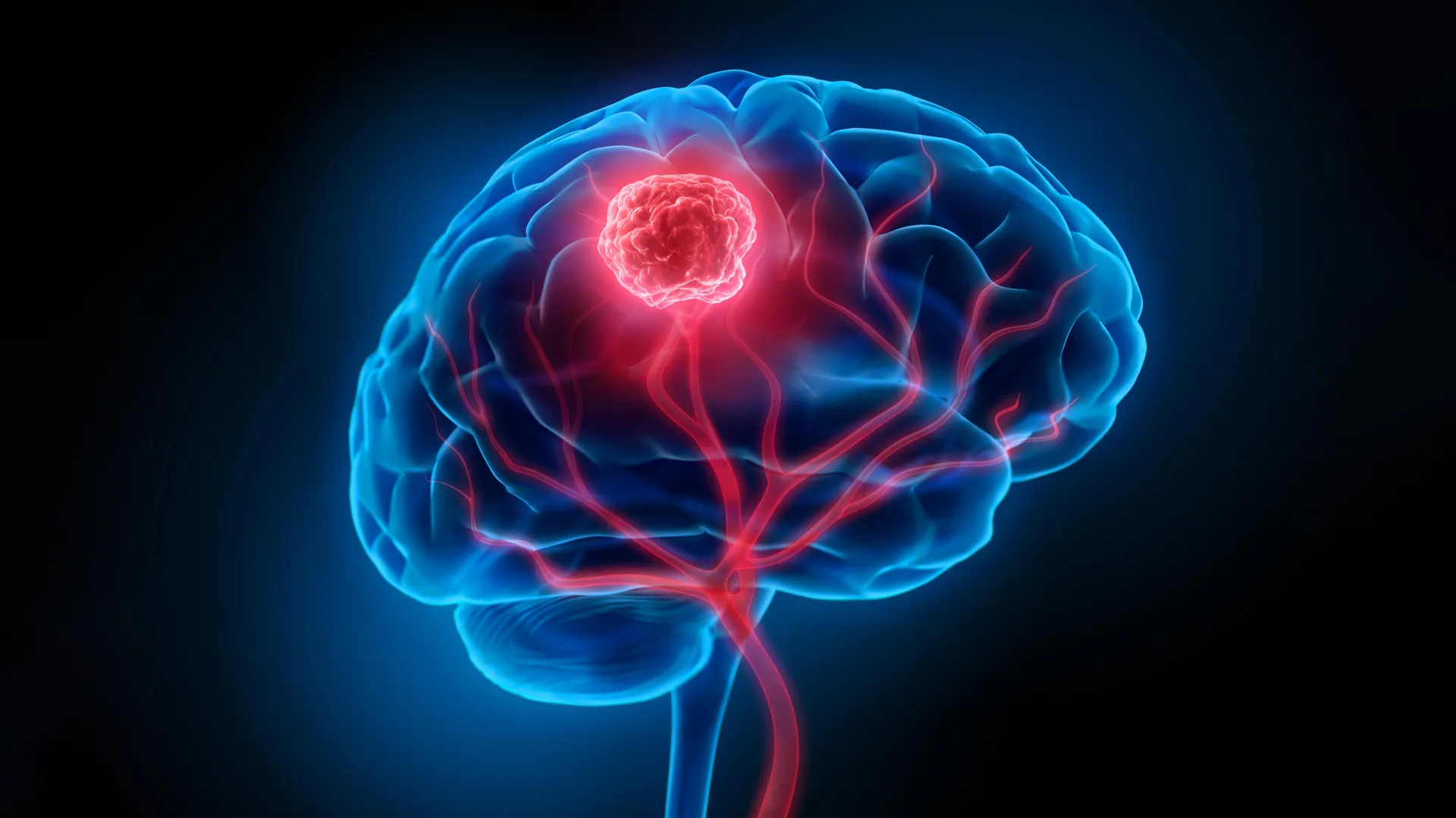
A groundbreaking investigation from South Korea has fundamentally reshaped our understanding of IDH-mutant glioma, the predominant form of aggressive brain cancer in adults under fifty, revealing that its insidious origins can precede detectable tumor formation by many years. Historically, therapeutic strategies have centered on the surgical excision of visible tumor masses identified through advanced imaging techniques, a reactive approach that often proves insufficient due to the cancer’s tenacious tendency to resurface post-treatment. However, this latest scientific endeavor, spearheaded by researchers at the Korea Advanced Institute of Science and Technology (KAIST) and Yonsei University Severance Hospital, illuminates a critical earlier phase of the disease: the silent proliferation of genetically altered brain cells long before they coalesce into a discernible malignancy.
This revelation stems from the meticulous identification of the cellular bedrock from which IDH-mutant gliomas emerge. The research team, under the joint leadership of Professor Jeong Ho Lee of KAIST’s Graduate School of Medical Science and Engineering and Professor Seok-Gu Kang of Yonsei University Severance Hospital, has pinpointed Glial Progenitor Cells (GPCs) – cells normally resident within healthy brain tissue – as the initial site of transformation. These GPCs, possessing the inherent capacity to differentiate into various glial cell types that support neuronal function, can, upon acquiring the specific IDH mutation, initiate a cascade of events that ultimately leads to cancerous growth. The significance of this finding lies in its challenge to the conventional view of tumor development, suggesting that the disease process is not a sudden event but rather a protracted, multi-stage evolution beginning at the cellular level.
The investigative methodology employed by the scientists involved a rigorous examination of both surgically resected tumor specimens and adjacent brain tissue that, to the unaided eye, appeared entirely normal. This comparative analysis yielded a pivotal observation: the presence of cells bearing the characteristic IDH mutation within regions of the brain that presented no macroscopic signs of abnormality. This suggests that the earliest manifestations of the cancer are not only microscopic but also geographically dispersed, a characteristic that profoundly complicates eradication efforts. The implications are substantial, offering a potential explanation for the high rates of recurrence observed in patients diagnosed with this type of glioma, as residual mutated cells, undetectable by current imaging, may lie dormant, awaiting an opportunity to proliferate.
To definitively characterize these nascent, mutated cells, the researchers leveraged cutting-edge "spatial transcriptomics." This sophisticated analytical technology allows for the simultaneous mapping of gene expression patterns within their precise anatomical locations. By applying this technique, the team unequivocally confirmed that the cells harboring the IDH mutation were, in fact, Glial Progenitor Cells situated within the cerebral cortex. This precision in identifying both the cell type and its location provides an unprecedented level of insight into the tumor’s inception. Furthermore, to validate their findings and recapitulate the developmental trajectory of the cancer, the researchers ingeniously engineered an animal model. By introducing the same genetic "driver mutation" observed in human patients into the GPCs of mice, they successfully replicated critical stages of brain tumor development, thereby providing robust experimental support for their hypothesis.
This latest research builds upon a foundational understanding established by the same research group in 2018, when they elucidated the origins of IDH wildtype glioblastoma, another aggressive brain malignancy. In that earlier study, the team identified neural stem cells residing in the subventricular zone – a critical niche for neurogenesis in the adult brain – as the precursor cells for this distinct subtype of glioma. The current findings underscore a crucial biological divergence: while both IDH wildtype glioblastoma and IDH-mutant glioma are classified as malignant brain tumors, they originate from fundamentally different cellular populations and initiate their developmental pathways in disparate locations within the brain. This stark contrast reinforces the concept that brain cancers, far from being monolithic entities, exhibit considerable heterogeneity in their origins and progression, dictated by their specific molecular subtypes.
The profound implications of this research extend directly to the realms of early diagnosis and therapeutic intervention. Professor Seok-Gu Kang, a co-corresponding author on the study, emphasized the paradigm-shifting nature of this discovery. He articulated that the understanding that brain tumors may not necessarily commence at the site of visible tumor mass necessitates a recalibration of clinical strategies. "A targeted approach focused on the origin cells and the site of origin according to the brain tumor subtype will serve as a crucial clue to changing the paradigm of early diagnosis and recurrence suppression treatment," he stated. This suggests that future diagnostic modalities might aim to detect the presence of these early-stage, mutated cells before the formation of a clinically apparent tumor, thereby enabling intervention at a far earlier, potentially more treatable, stage.
In anticipation of these advancements, Sovagen Co., Ltd., a KAIST faculty startup, is actively engaged in the development of novel RNA-based therapeutics. These innovative drugs are designed with the explicit goal of retarding or halting the progression and preventing the recurrence of IDH-mutant malignant brain tumors, capitalizing on the newly acquired knowledge of their cellular origins. Concurrently, Severance Hospital is pioneering the development of technologies aimed at the early detection and management of these nascent mutated cells, an initiative being pursued through the collaborative Korea-US Innovative Result Creation R&D project. These parallel efforts highlight a swift translation of fundamental scientific discovery into tangible clinical applications.
The genesis of this pivotal research can be traced back to a fundamental question posed by Dr. Jung Won Park, a postdoctoral researcher at KAIST’s Graduate School of Medical Science and Engineering and the study’s sole first author. A neurosurgeon by training, Dr. Park articulated the critical role of interdisciplinary collaboration, stating, "This achievement was made possible by combining KAIST’s world-class basic science research capabilities with the clinical expertise of Yonsei Severance Hospital." He further elaborated on the intellectual spark that ignited the investigation: "The question I kept asking while treating patients — ‘Where does this tumor originate?’ — was the starting point of this research." This dedication to unraveling the fundamental origins of disease, driven by clinical experience, exemplifies the synergy between basic science and clinical practice. The comprehensive findings of this collaborative effort were formally published on January 8th in the prestigious scientific journal Science, marking a significant contribution to the field of neuro-oncology. The research was generously supported by funding from the Suh Kyung-bae Science Foundation, the National Research Foundation of Korea, the Ministry of Science and ICT, the Ministry of Health and Welfare, and the Korea Health Industry Development Institute through its Physician-Scientist Training Program, underscoring the broad institutional commitment to advancing cancer research.