Scientists have illuminated a previously unrecognized biological mechanism integral to the healing of the central nervous system, offering a profound new avenue for therapeutic development in conditions ranging from spinal cord injuries to debilitating neurological disorders like multiple sclerosis and stroke. This groundbreaking research, detailed in the prestigious journal Nature, centers on a surprising and pivotal role played by astrocytes, a fundamental class of glial cells long recognized for their supportive functions within the brain and spinal cord. The investigation reveals that astrocytes situated at a considerable distance from an injury site are, in fact, key drivers of spinal cord repair. Furthermore, the study uncovers a sophisticated signaling pathway these specialized astrocytes employ to orchestrate the immune system’s critical task of clearing cellular debris, a prerequisite for effective tissue regeneration.
These newly identified cells have been designated "lesion-remote astrocytes," or LRAs, by the research team, who further delineated several distinct subtypes among them. For the first time, this research provides a comprehensive explanation of how one particular subtype of LRA can perceive damage occurring far from its location and initiate a cascade of responses that actively promote recovery. This discovery challenges existing paradigms of neural repair, which have often focused on immediate cellular responses at the injury site itself.
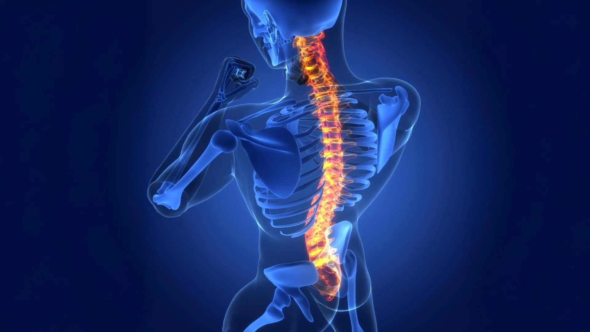
To understand the implications of these findings, it is essential to grasp the intricate architecture and response of the spinal cord to trauma. The spinal cord, a vital conduit of information between the brain and the body, comprises an inner core of gray matter, rich in neuronal cell bodies and astrocytes, surrounded by white matter. The white matter consists of myelinated nerve fibers, also ensheathed by astrocytes, which transmit electrochemical signals with remarkable speed and precision. Astrocytes within this structure are paramount in maintaining a tightly regulated microenvironment, ensuring the unimpeded flow of neural communication.
When the spinal cord sustains an injury, such as from impact or disease, the delicate network of nerve fibers is disrupted, leading to a cascade of cellular events. This tearing of nerve fibers results in the breakdown of tissue, generating fragments of cellular material that can impede further healing and neural function. In many tissues of the body, the inflammatory response, a natural defense mechanism, is typically localized to the site of damage. However, the elongated nature of neural pathways in the spinal cord means that damage and subsequent inflammation can propagate far beyond the initial point of impact, complicating the healing process and contributing to widespread functional deficits, including paralysis and sensory disturbances.
The Cedars-Sinai research team’s experiments, primarily conducted in murine models of spinal cord injury, unequivocally demonstrated the crucial role of LRAs in fostering repair processes. Crucially, the study also identified compelling evidence suggesting that this same cellular signaling mechanism is active in human spinal cord tissue affected by injury.
A specific subtype of LRA was found to produce a signaling protein known as CCN1. This molecule acts as a molecular messenger, communicating directly with immune cells within the central nervous system, specifically microglia. Microglia are the resident immune cells of the brain and spinal cord, performing essential housekeeping duties, including the removal of cellular debris. Following tissue damage, these microglia engage in phagocytosis, engulfing fragments of nerve fibers. However, nerve fiber debris is particularly rich in lipids, which can present a metabolic challenge for microglia, akin to indigestion, potentially hindering their efficiency. The study’s pivotal finding is that CCN1, secreted by LRAs, signals to these microglia, prompting a metabolic shift that enhances their ability to process and digest the fatty components of the debris.
This improved clearance of cellular detritus, facilitated by the LRA-microglia communication, may offer a crucial explanation for the partial, spontaneous recovery observed in some individuals following spinal cord injury. When the researchers experimentally inactivated the production of astrocyte-derived CCN1, the spinal cord’s healing capacity was significantly diminished. As explained by the study’s senior author, neuroscientist Joshua Burda, PhD, "If we remove astrocyte CCN1, the microglia eat, but they don’t digest. They call in more microglia, which also eat but don’t digest." This inefficient process leads to the accumulation of large aggregates of debris-laden microglia, exacerbating inflammation along the spinal cord and ultimately hindering tissue repair.
The implications of this discovery extend beyond acute spinal cord trauma. When examining spinal cord samples from individuals diagnosed with multiple sclerosis (MS), a chronic inflammatory disease that damages the myelin sheath surrounding nerve fibers, the researchers observed the identical CCN1-mediated repair process. This suggests that the fundamental principles of LRA-driven immune modulation and debris clearance may be broadly applicable to a range of neurological conditions affecting both the brain and the spinal cord.
David Underhill, PhD, Chair of the Department of Biomedical Sciences, underscored the significance of this underappreciated aspect of glial cell function, stating, "The role of astrocytes in central nervous system healing is remarkably understudied. This work strongly suggests that lesion-remote astrocytes offer a viable path for limiting chronic inflammation, enhancing functionally meaningful regeneration, and promoting neurological recovery after brain and spinal cord injury and in disease."
Dr. Burda and his team are now actively pursuing the development of therapeutic strategies aimed at harnessing the CCN1 signaling pathway to enhance spinal cord healing. Their ongoing research also explores the potential influence of astrocyte CCN1 on inflammatory neurodegenerative diseases and the aging process, further broadening the potential impact of this discovery. The collaborative effort behind this research included a substantial team of scientists from Cedars-Sinai, alongside researchers from other institutions, highlighting the interdisciplinary nature of modern scientific inquiry. The funding for this complex and vital research was provided by a consortium of prestigious organizations, including the National Institutes of Health (NIH), the Paralyzed Veterans Research Foundation of America, Wings for Life, the Cedars-Sinai Center for Neuroscience and Medicine, the American Academy of Neurology, the California Institute for Regenerative Medicine, the United States Department of Defense USAMRAA, and the Arnold O. Beckman Postdoctoral Fellowship, underscoring the significant investment in advancing our understanding of neurological repair.