A sophisticated artificial intelligence framework, dubbed CytoDiffusion, is poised to redefine the landscape of hematological diagnoses, offering a potent new tool for identifying subtle anomalies in blood cells that may elude even experienced human observation. This innovative system leverages generative artificial intelligence, a technology akin to that powering advanced image creation platforms, to meticulously scrutinize the intricate visual characteristics of blood cells. Instead of merely recognizing overt, predefined patterns, CytoDiffusion delves into the nuanced variations that characterize cellular appearance under microscopic examination, thereby enhancing diagnostic precision and consistency in conditions like leukemia.
The development of CytoDiffusion represents a significant departure from conventional approaches in medical AI, which often rely on classifying images into pre-established categories. The research team behind this breakthrough has demonstrated that their generative AI model possesses the capacity to comprehend the full spectrum of normal blood cell morphologies. This comprehensive understanding enables it to reliably detect and flag exceedingly rare or atypical cells that could signify the presence of underlying disease. This pioneering work, a collaborative effort involving researchers from the University of Cambridge, University College London, and Queen Mary University of London, has been formally documented and published in the esteemed journal, Nature Machine Intelligence.
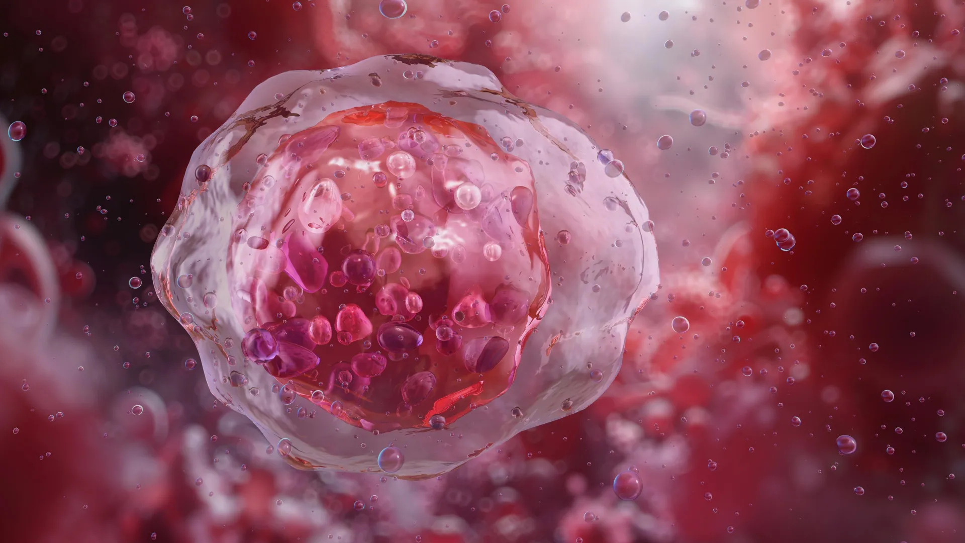
The accurate identification of minute discrepancies in the size, shape, and structural integrity of blood cells is fundamental to the diagnosis of a vast array of hematological disorders. However, cultivating the expertise required for such precise analysis is a protracted process, demanding years of dedicated training and experience. Even highly skilled hematologists may encounter situations where their interpretations of complex cases diverge, introducing an element of diagnostic uncertainty. Dr. Simon Deltadahl, the study’s lead author and a researcher at Cambridge’s Department of Applied Mathematics and Theoretical Physics, explained the inherent complexity, stating, "We possess a multitude of blood cell types, each with distinct properties and vital functions within our bodies. For instance, white blood cells are specialized in combating infections. Nevertheless, discerning the appearance of an unusual or diseased blood cell under a microscope is a critical component in diagnosing numerous illnesses."
The sheer volume of cells present in a standard blood smear presents a formidable challenge for manual examination. A single blood smear can contain tens of thousands of individual cells, an amount far exceeding what any single human can realistically scrutinize on a cell-by-cell basis. "It is simply not feasible for humans to examine every cell in a smear," Dr. Deltadahl elaborated. "Our model automates this laborious task, efficiently triaging routine samples and flagging any irregularities for expert human review." This sentiment is echoed by clinicians who have grappled with this diagnostic bottleneck. Dr. Suthesh Sivapalaratnam, a co-senior author from Queen Mary University of London and a former junior hematology doctor, shared his personal experience: "As a junior hematology doctor, a significant clinical challenge I faced was the substantial volume of blood films requiring analysis at the end of each workday. While I was diligently examining them in the late hours, I became increasingly convinced that an AI system would ultimately perform this task more effectively than I could."
The foundation of CytoDiffusion’s capabilities lies in its training on an unprecedentedly large dataset. The researchers meticulously curated and utilized over half a million blood smear images, amassed from Addenbrooke’s Hospital in Cambridge. This extensive collection, recognized as the most comprehensive of its kind, encompasses a wide array of common blood cell types, alongside rare and unusual examples, and crucially, includes cellular features that frequently pose difficulties for existing automated diagnostic systems.
A key differentiator of CytoDiffusion’s training methodology is its focus on modeling the entire generative distribution of blood cell appearances, rather than simply learning to categorize cells into discrete, predefined classes. This holistic approach imbues the AI with enhanced resilience to variations introduced by different hospitals, microscopes, and staining techniques. Furthermore, it significantly bolsters its proficiency in identifying rare or aberrant cells.
During rigorous testing, CytoDiffusion demonstrated a marked improvement in detecting abnormal cells associated with leukemia, exhibiting substantially higher sensitivity compared to existing automated systems. Its performance was on par with, or even surpassed, that of leading current models, even when trained with considerably fewer examples. A notable advancement is the system’s ability to quantify its own level of confidence in its predictions, a crucial aspect for clinical decision-making. "When we evaluated its accuracy, the system performed slightly better than humans," Dr. Deltadahl noted. "However, its true distinction emerged in its awareness of uncertainty. Our model would never confidently misidentify a cell; whereas, this is a fallibility that humans can occasionally exhibit."
Professor Michael Roberts, a co-senior author from Cambridge’s Department of Applied Mathematics and Theoretical Physics, highlighted the system’s evaluation against real-world diagnostic challenges: "We assessed our method against numerous obstacles commonly encountered in practical AI applications, such as processing novel images, images acquired by diverse instrumentation, and the inherent ambiguity in data labels. This comprehensive framework provides a multifaceted perspective on model performance, which we believe will prove invaluable to the broader research community."
Intriguingly, the research team also discovered CytoDiffusion’s capacity to generate synthetic blood cell images that are virtually indistinguishable from authentic microscopic specimens. In a sophisticated ‘Turing test’ involving ten seasoned hematologists, the specialists were unable to reliably differentiate between genuine blood cell images and those fabricated by the AI, performing no better than random chance. "This outcome was truly astonishing," Dr. Deltadahl confessed. "These are individuals who dedicate their professional lives to scrutinizing blood cells, and even they could not discern the artificial from the real."
In a move to foster global scientific advancement, the researchers are making accessible what they describe as the world’s largest publicly available repository of peripheral blood smear images, comprising more than half a million individual samples. "By providing this resource openly, our objective is to empower researchers worldwide to develop and rigorously test novel AI models," Dr. Deltadahl stated. "This initiative aims to democratize access to high-quality medical data, ultimately contributing to enhanced patient care on a global scale."
Despite the overwhelmingly positive findings, the research team is emphatic in their assertion that CytoDiffusion is not designed to supplant human clinicians. Instead, its primary role is to serve as an invaluable assistant, empowering medical professionals by rapidly identifying cases that warrant immediate attention and by efficiently processing routine diagnostic samples. Professor Parashkev Nachev, a co-senior author from UCL, articulated the broader vision for healthcare AI: "The genuine value of AI in healthcare lies not in replicating human expertise at a reduced cost, but in augmenting diagnostic, prognostic, and prescriptive capabilities beyond what individual experts or simple statistical models can achieve. Our research suggests that generative AI will be central to this mission, revolutionizing not only the precision of clinical support systems but also their insight into their own limitations. This ‘metacognitive’ awareness – the ability to recognize the boundaries of one’s own knowledge – is paramount in clinical decision-making, and our findings indicate that machines may possess this attribute to a greater degree than humans."
The researchers acknowledge that further investigation is necessary to optimize the system’s processing speed and to validate its performance across a more diverse range of patient demographics, ensuring its accuracy and equitable application. This comprehensive study benefited from the support of various esteemed organizations, including The Trinity Challenge, Wellcome, the British Heart Foundation, Cambridge University Hospitals NHS Trust, Barts Health NHS Trust, the NIHR Cambridge Biomedical Research Centre, NIHR UCLH Biomedical Research Centre, and NHS Blood and Transplant. The research was conducted under the auspices of the Imaging working group within the BloodCounts! consortium, an initiative dedicated to advancing blood diagnostics globally through the application of AI. Dr. Simon Deltadahl is affiliated with Lucy Cavendish College, Cambridge.