A groundbreaking advancement in medical diagnostics has emerged from a collaborative effort between researchers at the California Institute of Technology (Caltech) and the University of Southern California (USC), introducing a novel imaging methodology capable of generating comprehensive three-dimensional color representations of internal body structures. This innovative system, known as Rotational Ultrasound and Photoacoustic Tomography (RUS-PAT), uniquely combines the strengths of conventional ultrasound with photoacoustic imaging, offering an unprecedented dual perspective on both the anatomical composition of soft tissues and the intricate functional dynamics of blood vessels. Initial demonstrations highlight the technique’s versatility across various anatomical regions, holding substantial promise for enhancing early disease detection, refining diagnostic precision, and advancing fundamental biological research, particularly in areas such as breast cancer detection, monitoring diabetic neuropathy, and elucidating brain function. The detailed scientific underpinnings and experimental validations of this work were formally presented in the esteemed journal Nature Biomedical Engineering.
For decades, medical practitioners and researchers have relied on a suite of imaging technologies, each with distinct advantages and inherent compromises. The quest for a single, non-invasive tool that could simultaneously provide detailed structural anatomy and critical physiological information without significant drawbacks has been a persistent challenge. Standard ultrasonography, for instance, remains a cornerstone of diagnostic imaging due to its cost-effectiveness, portability, and real-time capabilities. It operates by emitting high-frequency sound waves into the body and interpreting the echoes reflected from various tissues. While excellent for visualizing the shape, size, and location of organs and detecting gross structural abnormalities in two dimensions, its ability to differentiate subtle tissue characteristics or track dynamic physiological processes like blood flow with high specificity is often limited. Furthermore, its field of view can be narrow, requiring skilled operators to scan and mentally reconstruct a complete picture.
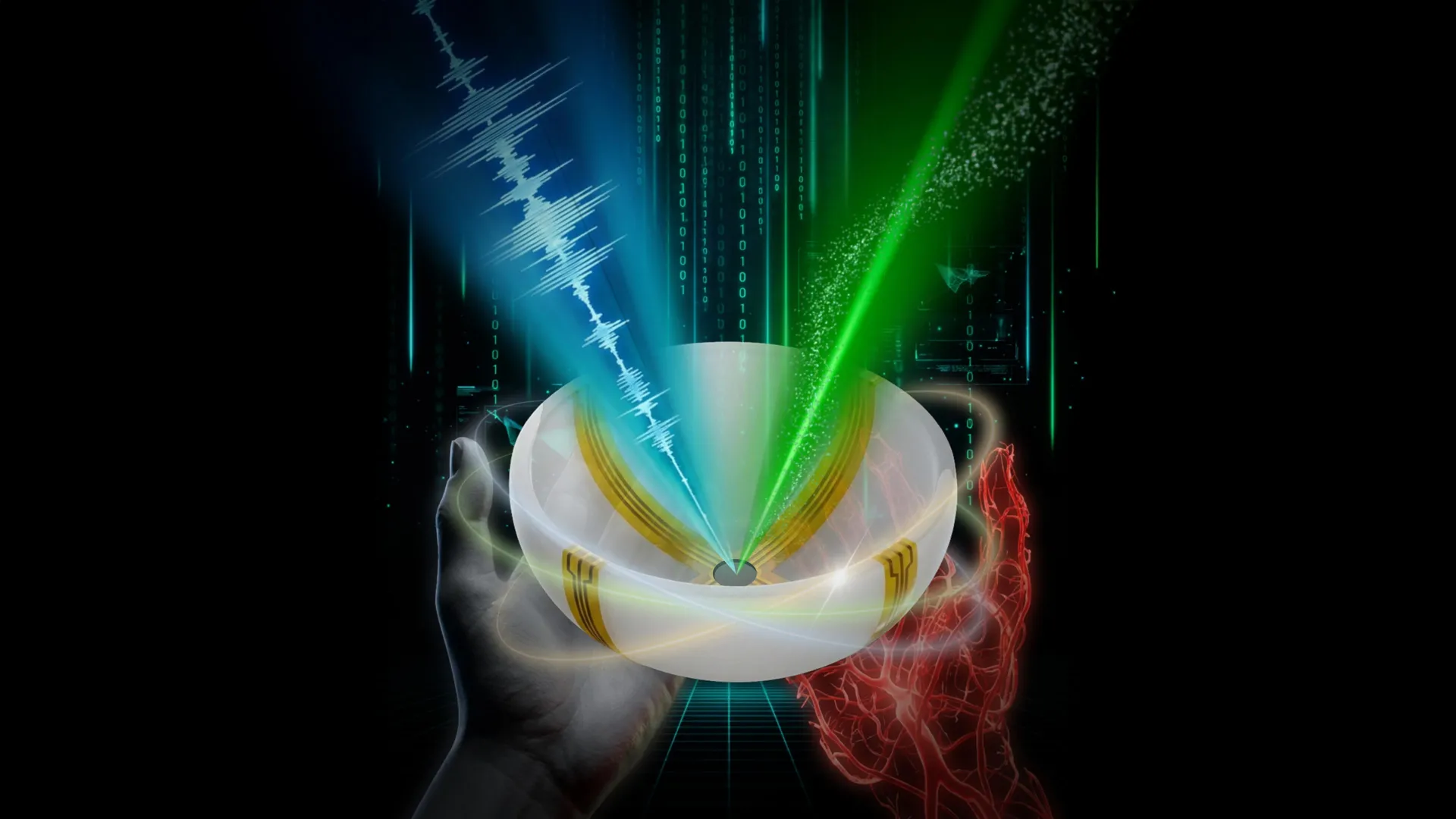
Complementing ultrasound, photoacoustic imaging (PAT) provides a different, yet crucial, dimension of information. This technique leverages the photoacoustic effect, where short pulses of laser light are absorbed by specific molecules within tissues, particularly hemoglobin in red blood cells. This absorption causes transient thermal expansion, generating localized ultrasound waves that are then detected by transducers. The resulting signals are used to construct images that vividly display blood vessels in optical color, offering a direct visualization of vascular networks and quantifiable insights into blood oxygenation and flow patterns. This molecular-level information is invaluable for identifying areas of increased metabolic activity, such as those often associated with tumor growth or inflammation. However, a significant drawback of PAT has been its relative inability to resolve fine anatomical details of the surrounding tissue, leaving a gap in contextual understanding.
Other widely used diagnostic modalities, such as Computed Tomography (CT) and Magnetic Resonance Imaging (MRI), address some of these limitations but introduce their own set of tradeoffs. CT scans, which employ X-rays, excel at providing highly detailed cross-sectional images of bones and soft tissues, but they expose patients to ionizing radiation, a concern for repeated or frequent examinations. MRI, utilizing powerful magnetic fields and radio waves, offers superior soft tissue contrast without radiation exposure, making it invaluable for neurological, musculoskeletal, and oncological imaging. Nevertheless, MRI procedures are often lengthy, expensive, and can be challenging for patients with claustrophobia or certain metallic implants. Both CT and MRI frequently necessitate the administration of contrast agents, which, while generally safe, carry potential risks for allergic reactions or kidney complications in some individuals. The cumulative effect of these limitations across existing technologies underscored a critical unmet need for an imaging platform that could deliver comprehensive structural and functional data efficiently, safely, and affordably.
The genesis of RUS-PAT lies in the visionary work of Lihong Wang, the Bren Professor of Medical Engineering and Electrical Engineering and the Andrew and Peggy Cherng Medical Engineering Leadership Chair at Caltech, who pioneered photoacoustic tomography over two decades ago. Recognizing the complementary strengths of ultrasound and photoacoustic imaging, Professor Wang spearheaded the ambitious project to integrate these two powerful modalities. His goal was not merely to combine them additively, but to forge an optimal synergy where the combined system would surmount the individual weaknesses of each technique. This aspiration led to the development of RUS-PAT, a system designed to concurrently capture both the anatomical architecture provided by ultrasound and the molecular-level functional insights offered by photoacoustics.
The engineering challenge in merging these systems was considerable. Traditional ultrasound imaging relies on arrays of transducers that both emit and receive sound waves, creating complex and often costly hardware. Direct integration of such an elaborate ultrasound system with photoacoustic imaging, which primarily requires sophisticated detection of light-induced sound waves, seemed unwieldy and impractical for widespread clinical adoption. Professor Wang’s pivotal insight was to re-evaluate the fundamental requirements of each modality. He realized that while ultrasound needed both emission and reception, photoacoustic imaging primarily necessitated robust detection. This led him to conceptualize a simpler, more elegant solution: could a single, wide-field ultrasound transducer be adapted to both generate the sound waves for structural imaging and simultaneously detect all acoustic signals, including those produced by laser-induced photoacoustic effects?
This conceptual leap materialized into a revolutionary system design. RUS-PAT employs a limited number of arc-shaped ultrasound detectors strategically positioned to rotate around the subject. This rotational mechanism effectively mimics the comprehensive data acquisition capabilities of a much larger, more complex hemispheric array, but with significantly fewer components. By utilizing a single transducer for both emitting ultrasound waves (for anatomical imaging) and receiving the resulting echoes, as well as the photoacoustically generated sound waves, the system dramatically simplifies hardware requirements. This innovative architecture not only reduces manufacturing complexity and cost but also facilitates a more streamlined data acquisition process, paving the way for its practical application in diverse clinical settings.
The potential clinical ramifications of RUS-PAT are profound and far-reaching, as highlighted by Dr. Charles Y. Liu, a co-author of the study and a visiting associate in biology and biological engineering at Caltech, who also holds professorships at the Keck School of Medicine of USC and directs USC’s Neurorestoration Center. Dr. Liu emphasized that the novel fusion of acoustic and photoacoustic techniques directly addresses many critical limitations inherent in current widely adopted medical imaging practices. Crucially, the system’s feasibility for human application has already been unequivocally demonstrated in multiple contexts, signaling its readiness for further progression toward clinical integration.
One of the most significant anticipated applications is in the realm of breast cancer imaging. Current diagnostic pathways often involve mammography for initial screening, followed by ultrasound or MRI for further characterization. While these methods are effective, they sometimes struggle to differentiate benign from malignant lesions based solely on structural characteristics or may require biopsies for definitive diagnosis. RUS-PAT offers a transformative advantage by providing simultaneous structural information (tumor size, shape, borders) alongside functional insights into its biological activity (blood vessel density, oxygenation, and angiogenesis—the formation of new blood vessels that feed tumors). This combined information could enable earlier and more accurate identification of suspicious lesions, better characterization of tumor aggressiveness, and improved guidance for biopsies or treatment planning, potentially reducing unnecessary interventions and enhancing patient outcomes.
For patients grappling with diabetic neuropathy, a debilitating complication of diabetes that affects nerve function and blood supply, RUS-PAT presents a groundbreaking monitoring tool. Diabetic neuropathy often involves damage to peripheral nerves and compromise of the microvasculature supplying these nerves. Existing methods for tracking this condition are often limited in their ability to simultaneously assess both nerve structural integrity and the critical oxygen supply to these tissues. With RUS-PAT, physicians could non-invasively monitor both the anatomical changes in nerve tissue and the corresponding microvascular perfusion in a single, rapid scan. This comprehensive view could facilitate earlier detection of disease progression, enable more precise assessment of treatment efficacy, and ultimately contribute to better management strategies for preventing irreversible nerve damage.
Beyond specific disease applications, Professor Wang also underscored RUS-PAT’s immense potential for advancing fundamental brain research. The brain’s intricate structure and dynamic functional activity, particularly the relationship between neural activity and blood flow (neurovascular coupling), are areas of intense scientific inquiry. Traditional methods for studying brain anatomy and function often require separate imaging sessions or specialized equipment. RUS-PAT could allow scientists to simultaneously visualize detailed brain anatomy and observe real-time blood flow dynamics within specific brain regions. This capability could unlock new avenues for understanding neurological disorders like stroke, Alzheimer’s disease, or even the basic mechanisms of learning and memory by providing a comprehensive, integrated view of the brain’s form and function.
The practical advantages of RUS-PAT extend to its operational characteristics. Each scan is remarkably swift, completing in less than one minute, which significantly improves patient throughput and comfort compared to lengthier procedures. Currently, the system can image tissues up to approximately 4 centimeters deep, a depth suitable for many superficial structures and organs. However, the modular nature of light delivery, including the potential integration with endoscopic tools, offers a promising pathway to access deeper anatomical regions within the body, such as the gastrointestinal tract or arterial lumens. The system’s design places ultrasound transducers and a laser beneath a scanning bed, facilitating ease of use. Importantly, early testing phases have already involved human volunteers and patients, demonstrating the technology’s safety and efficacy in a real-world context and marking its initial steps toward broader clinical deployment.
The detailed findings of this innovative research were published under the title "Rotational ultrasound and photoacoustic tomography of the human body." The collaborative effort was spearheaded by co-lead authors Yang Zhang, Shuai Na, and Dr. Jonathan J. Russin. Zhang and Na conducted their pivotal work as postdoctoral researchers at Caltech and have since transitioned to Tsinghua University and Peking University in Beijing, respectively. Dr. Russin is affiliated with the Keck School of Medicine of USC and the Rancho Los Amigos National Rehabilitation Center in Downey, California. Additional significant contributions were made by a team of Caltech researchers, including Karteekeya Sastry, Li Lin, Junfu Zheng, Yilin Luo, Xin Tong, Yujin An, Peng Hu, and former research scientist Konstantin Maslov, with Li Lin now based at Zhejiang University in Hangzhou, China. Dr. Tze-Woei Tan from the Keck School of Medicine of USC also served as a co-author. This groundbreaking research received critical financial backing from the National Institutes of Health, underscoring its recognized importance to public health and scientific advancement. As RUS-PAT continues its journey from laboratory innovation to clinical application, it promises to reshape the landscape of medical imaging, offering a more holistic and insightful window into the complexities of human health and disease.