A breakthrough in biomedical optics has yielded a highly refined imaging apparatus capable of discerning cancerous tissue from healthy cells with exceptional accuracy, potentially ushering in an era of earlier cancer diagnosis and facilitating the integration of sophisticated molecular visualization techniques into routine medical practice. This innovative system, developed by a team of researchers, promises to bridge the gap between cutting-edge laboratory discoveries and their practical application in clinical environments.
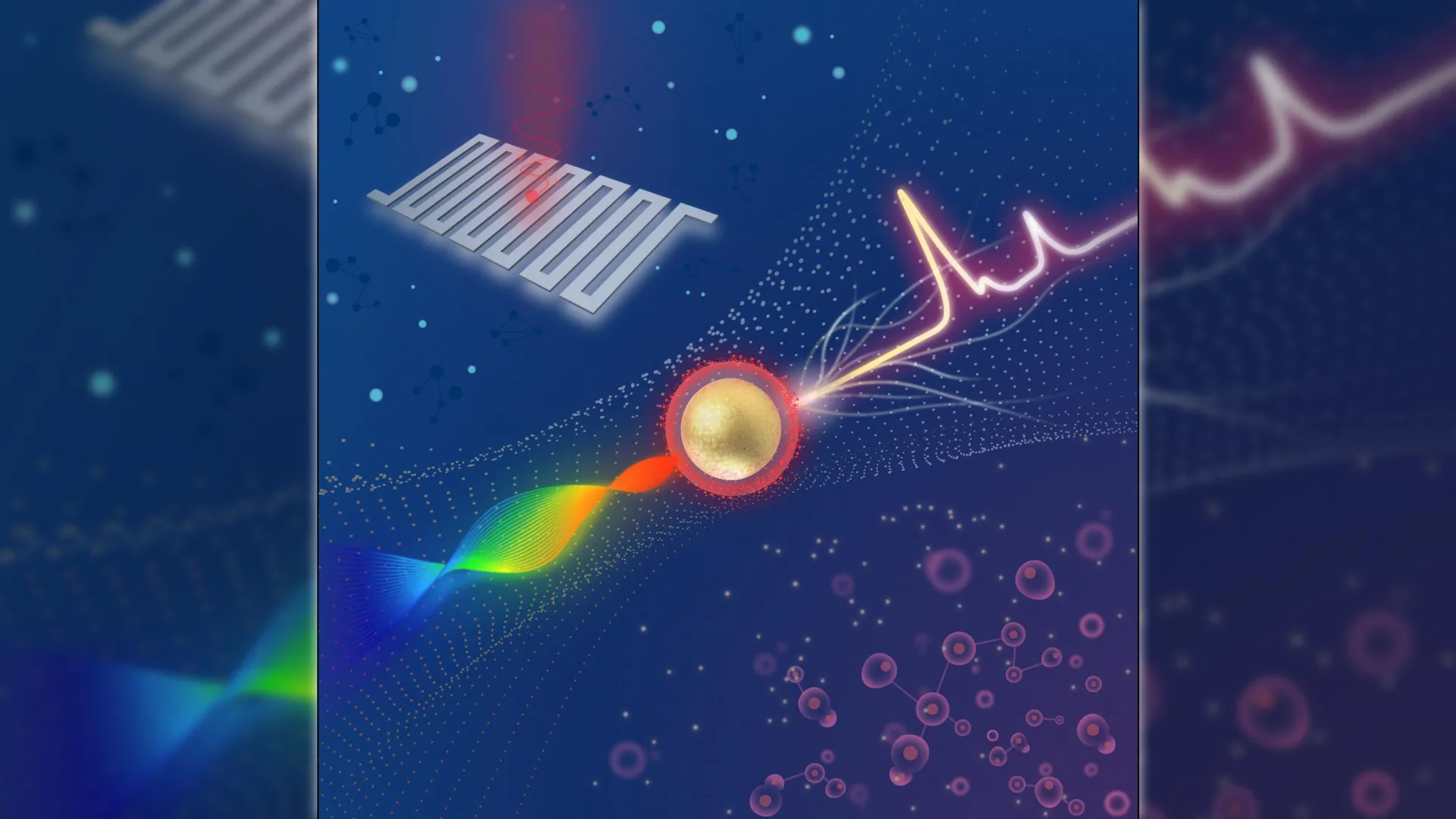
At the heart of this advancement lies a compact Raman imaging system meticulously engineered to detect the exceptionally faint signatures emitted by surface-enhanced Raman scattering (SERS) nanoparticles. These specially designed nanoparticles are programmed to selectively adhere to specific molecular indicators present on tumor cells. Once administered to a biological sample or introduced into the examination area, the system meticulously analyzes the Raman signals emanating from these nanoparticles, automatically flagging areas that exhibit a heightened probability of containing malignant tissue. This capability represents a significant departure from conventional diagnostic methodologies, which are often characterized by lengthy processing times and a heavy reliance on manual interpretation by skilled pathologists.
The research, published in the esteemed journal Optica, details a remarkable leap in sensitivity, with the new system demonstrating an ability to differentiate between cancerous and healthy cells by detecting Raman signals that are approximately four times weaker than those measurable by existing commercial systems. This amplified sensitivity is the result of a synergistic integration of a swept-source laser, which dynamically adjusts its wavelength during the analytical process, and an ultra-sensitive detector known as a superconducting nanowire single-photon detector (SNSPD). The SNSPD, a marvel of quantum technology, is capable of registering individual photons, thereby enabling the system to capture exceedingly faint optical signals with remarkable speed and an exceptionally low level of background interference.
Dr. Zhen Qiu, the lead investigator from Michigan State University’s Institute for Quantitative Health Science and Engineering (IQ), highlighted the limitations of current diagnostic methods, noting their inherent time consumption and labor-intensive nature, which typically involve the meticulous staining of tissue samples followed by careful microscopic examination by pathologists. He posited that while this new system is not intended to supplant traditional pathology outright, it holds immense potential as a rapid screening tool to expedite the diagnostic workflow. The ultimate vision for this technology, as articulated by Dr. Qiu, extends to the development of portable or even intraoperative devices that would empower clinicians to identify cancers at their nascent stages, refine the precision of biopsy sampling, and monitor disease progression through less invasive means. Such advancements, he anticipates, could profoundly improve patient outcomes by minimizing diagnostic delays and accelerating the transition from detection to therapeutic intervention.
The development of the SNSPD is a critical enabler of this imaging system’s enhanced performance. Dr. Qiu’s research group has been at the forefront of exploring the application of SNSPDs to augment a diverse array of imaging technologies. These detectors operate on the principle of a superconducting wire that is exquisitely sensitive to individual light particles. This sensitivity allows the system to acquire extremely weak optical signals at high velocities while simultaneously minimizing unwanted noise.
For this particular project, the research team set out to construct a platform capable of measuring Raman signals that are substantially fainter than those detectable by contemporary Raman systems. Raman imaging fundamentally operates by mapping the chemical composition of a sample through the unique light-scattering characteristics, or "fingerprints," of its constituent molecules. The intensity of these signals can be significantly amplified through the strategic utilization of SERS nanoparticles.
The integration of this advanced detector with a swept-source Raman architecture, which notably eschews the need for a bulky camera and achieves superior light collection efficiency, has culminated in a system that surpasses the detection limits of comparable commercial instruments. Furthermore, the system’s fiber coupling configuration and its inherently compact design are key factors that facilitate miniaturization and pave the way for its future deployment in clinical settings.
To rigorously evaluate the capabilities of the newly developed system, the researchers employed SERS nanoparticles functionalized with hyaluronic acid. This specific coating promotes the nanoparticles’ binding to CD44, a surface protein frequently overexpressed on various types of tumor cells. Initial investigations, conducted with straightforward nanoparticle solutions, demonstrated the system’s impressive sensitivity, reaching femtomolar levels. Subsequently, the imaging platform was applied to more complex biological samples, including cultured breast cancer cells, surgically removed mouse tumors, and healthy tissue specimens.
The experimental results unequivocally indicated that the SERS signals were markedly concentrated within the tumor samples, with only negligible background signals detected in the healthy tissue. This observation serves as a powerful testament to both the system’s exceptional sensitivity and its inherent ability to reliably differentiate between tumorous and healthy tissue. Crucially, the researchers noted that by modifying or substituting the targeting molecule, this innovative methodology could be readily adapted to detect a wide spectrum of other cancer types, underscoring its versatility.
Despite these promising findings, the researchers acknowledge that further development is imperative before the system can be widely adopted in clinical practice. Future research efforts will be directed towards enhancing the readout speed of the system and expanding the scope of validation studies. The team is actively exploring the potential of faster laser sources, including Vertical-Cavity Surface-Emitting Lasers (VCSELs), and investigating whether narrowing the laser’s wavelength sweep range can further optimize performance. Moreover, plans are underway to conduct multiplexing experiments, which involve the simultaneous use of different types of nanoparticles designed to target multiple biomarkers, thereby providing a more comprehensive molecular profile.
The research team expressed their gratitude to industry collaborator Quantum Opus, whose contribution in providing the SNSPD devices was instrumental to the successful execution of this work.