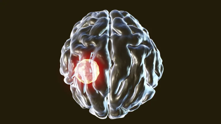
For generations, the prevailing dogma in neuroscience asserted a stark limitation: once damaged or destroyed, the intricate network of neurons within the brain and its sensory pathways possessed a near-absolute inability to regenerate. This fundamental tenet profoundly influenced the scientific community’s understanding of neurological trauma and dictated the therapeutic strategies available for recovery. However, observable phenomena – the frequent, albeit partial, restoration of lost functions following brain injury – persistently challenged this rigid doctrine, prompting a critical re-evaluation of the mechanisms underpinning recovery. The central enigma revolved around how functional rehabilitation could occur if the primary cellular components, neurons, were incapable of self-repair or replacement.
Recent groundbreaking research, published in the esteemed journal JNeurosci, has begun to unravel this complex puzzle, offering compelling new insights into the adaptive capabilities of the nervous system. A team led by Athanasios Alexandris at Johns Hopkins University employed a sophisticated experimental model in mice to meticulously investigate the intricate processes unfolding within the visual system subsequent to traumatic brain injury. The visual system, a marvel of biological engineering, encompasses a sophisticated chain of communication, beginning with photoreceptor cells in the retina of the eye that capture light and convert it into electrical signals, which are then transmitted via specialized neurons to various regions of the brain responsible for visual processing. Damage occurring anywhere along this critical pathway, from the eye to the cortical areas, can lead to significant disruptions in visual perception, ranging from blurred vision to complete blindness.
The investigators focused their attention on the intricate synaptic connections that bridge the gap between the retinal ganglion cells in the eye and their neuronal targets within the brain. Contrary to the expectation of widespread generation of new neurons – a process known as neurogenesis – the researchers observed a remarkably different, yet equally significant, phenomenon. Instead of relying on the birth of novel cells, the neurons that managed to survive the initial insult demonstrated an astonishing capacity for adaptation and remodeling.
These resilient, surviving neurons initiated a process of remarkable structural plasticity. They began to extend new, exploratory branches, or neurites, from their existing cellular bodies and axons. This dynamic outgrowth allowed these surviving cells to establish a greater number of connections with a broader network of neurons in the brain than they had prior to the injury. This adaptive mechanism, termed axonal sprouting, served as a critical compensatory strategy, effectively filling the functional void left by the neurons that had succumbed to the trauma. Over a sustained period, the study documented a significant restoration in the overall number of functional connections between the eye and the brain, with these re-established pathways approaching, and in some cases, even matching the density observed in uninjured control subjects.
Crucially, the observed restoration was not merely a superficial, structural reconstruction; it represented a genuine functional recovery. Electrophysiological recordings, which measure the electrical activity of neurons, provided compelling evidence that these newly formed or re-established neural circuits were not only structurally sound but also actively transmitting neural signals with remarkable fidelity. This functional integration meant that the visual system was regaining its capacity to process and transmit information, thereby enabling a return of visual perception, despite the presence of significant underlying damage. This finding has profound implications, suggesting that the brain’s inherent resilience and ability to reorganize itself play a far more substantial role in recovery than previously appreciated.
Adding another layer of complexity and significance to these findings, the research unearthed a striking sex-dependent difference in the efficacy of this visual system recovery process. While male mice exhibited a robust and effective recovery, largely driven by the compensatory sprouting mechanism described, female mice demonstrated a markedly slower or, in some instances, incomplete repair. The regeneration of eye-to-brain connections in the female subjects did not consistently reach the pre-injury levels observed in the males.
The authors posit that these observed disparities underscore the existence of distinct recovery mechanisms that are modulated by sex. As Alexandris himself elaborated, the discovery of these sex differences was unanticipated, yet it resonates strongly with anecdotal and clinical observations in human populations. It is widely recognized that women often report a higher incidence of persistent post-concussion symptoms and longer recovery times following traumatic brain injuries compared to men. The researchers’ ongoing work aims to elucidate the precise molecular and cellular underpinnings of this observed branch sprouting phenomenon, and critically, to identify the factors that may impede or delay this restorative process in females. Understanding these sex-specific biological pathways could pave the way for the development of targeted therapeutic interventions designed to enhance neural recovery across a spectrum of injuries, including concussions and other forms of neurological trauma.
The research team is committed to further exploring the biological factors that contribute to these sex-based variations in neural repair. By delving deeper into the molecular signaling pathways, genetic predispositions, and hormonal influences that govern neuronal plasticity and compensatory sprouting, they aspire to uncover novel strategies that could significantly improve the prospects for healing and functional restoration following brain injuries. This research not only challenges long-held beliefs about neuronal regeneration but also opens promising new avenues for understanding and treating the devastating consequences of neurological damage.