Groundbreaking research emanating from neuroscientists at Trinity College Dublin has illuminated a remarkable cognitive milestone in human development, revealing that infants merely two months old possess the capacity to segment their visual environment into discernible object categories. This finding significantly challenges previous assumptions regarding the timeline of perceptual development, suggesting that the fundamental architecture for understanding the world is established exceptionally early in infancy, far sooner than had been heretofore understood. The implications of this research are profound, offering a deeper understanding of the nascent stages of human cognition and the intricate processes that underpin how we learn to perceive and interact with our surroundings from the very outset of life.
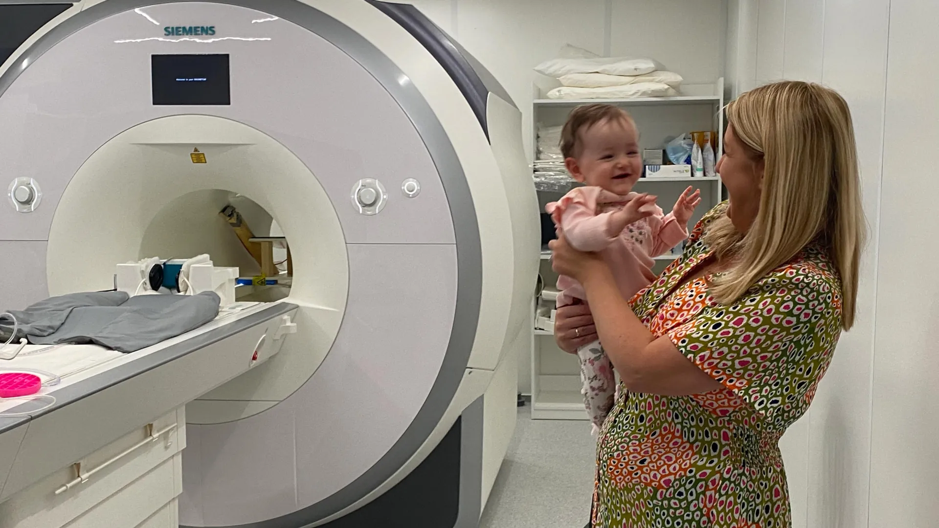
The study, meticulously detailed in the esteemed journal Nature Neuroscience, was undertaken by a dedicated team affiliated with the Trinity College Institute of Neuroscience (TCIN) and the School of Psychology. By ingeniously integrating advanced brain imaging techniques with sophisticated artificial intelligence models, the researchers were able to achieve an unprecedented window into the cognitive landscapes of very young infants. This innovative methodology allowed for the observation and analysis of neural activity during a period when verbal communication and deliberate motor control are virtually nonexistent, providing critical insights into the underlying mechanisms of infant thought and learning during these formative months. The collective efforts of the FOUNDCOG research team, in collaboration with the Coombe and Rotunda Hospitals in Dublin, culminated in the recruitment of 130 two-month-old participants. These infants were comfortably situated on soft beanbag chairs, equipped with sound-canceling headphones, and presented with a sequence of vibrant, engaging images designed to capture and maintain their attention for periods of 15 to 20 minutes.
This carefully controlled experimental environment facilitated the application of functional magnetic resonance imaging (fMRI) to meticulously record the intricate patterns of brain activity as the infants observed a diverse array of images. These visual stimuli were deliberately chosen to represent 12 distinct and familiar object categories, including common items such as cats, birds, rubber ducks, shopping carts, and trees. The objective was to ascertain whether the infant brain could begin to form conceptual groupings of these visually disparate objects, a task that requires a degree of abstract representation and categorization.
Following the acquisition of the brain scan data, the research team deployed powerful artificial intelligence algorithms to meticulously analyze how these different visual categories were encoded and represented within the developing infant brain. By drawing parallels between the neural activity patterns observed along established visual recognition pathways in both the AI models and the participants’ brains, the researchers were able to achieve a more nuanced comprehension of the nascent processes of categorization in early infancy. This comparative analysis proved instrumental in deciphering the complex neural signatures associated with object recognition and classification at such a tender age.
Dr. Cliona O’Doherty, the principal investigator of this groundbreaking study, who conducted the research while a member of Trinity’s Cusack Lab, articulated the enduring fascination surrounding the infant mind. "Parents and scientists have long wondered what goes on in a baby’s mind and what they actually see when they view the world around them," she stated, underscoring the profound implications of their findings. "This research highlights the richness of brain function in the first year of life." Dr. O’Doherty further elaborated on the significance of their discoveries, noting, "Although at two months, infants’ communication is limited by a lack of language and fine motor control, their minds were already not only representing how things look, but figuring out to which category they belonged. This shows that the foundations of visual cognition are already in place from very early on and much earlier than expected." This assertion points to a remarkably sophisticated level of cognitive processing occurring within the infant brain, operating on principles of abstract representation and classification well before the emergence of more overt cognitive and communicative abilities.
The pioneering nature of this study is further emphasized by Rhodri Cusack, the team leader and Thomas Mitchell Professor of Cognitive Neuroscience at Trinity’s School of Psychology and TCIN. He highlighted the study’s distinction as "the largest longitudinal study with functional magnetic resonance imaging (fMRI) of awake infants." Cusack explained that "The rich dataset capturing brain activity opens up a whole new way to measure what babies are thinking at a very early age. It also highlights the potential for neuroimaging and computational models to be used as a diagnostic tool in very young infants." The implications for early diagnostic capabilities are substantial, potentially enabling the identification of developmental anomalies or neurological differences at a critical juncture for intervention.
Beyond its immediate scientific contributions, the research holds significant promise for advancements in artificial intelligence. Cusack observed, "Babies learn much more quickly than today’s AI models and by studying how they do this, we hope to inspire a new generation of AI models that learn more efficiently, so reducing their economic and environmental costs." This interdisciplinary synergy, drawing inspiration from biological learning mechanisms to enhance technological capabilities, represents a forward-thinking approach to AI development, aiming for more sustainable and effective learning paradigms.
Dr. Anna Truzzi, now a Senior Lecturer in the School of Psychology at Queen’s University Belfast and a co-author of the paper, underscored the role of recent technological advancements in making this research feasible. "Until recently, we could not reliably measure how specific areas of the infant brain interpreted visual information," she commented. "By combining AI and neuroimaging, our study offers a very unique insight, which helps us to understand much more about how babies learn in their first year of life." Truzzi further elaborated on the critical period of early development, stating, "The first year is a period of rapid and intricate brain development. This study provides new foundational knowledge which will help guide early-years education, inform clinical support for neurodevelopmental conditions and inspire more biologically-grounded approaches in artificial intelligence." This positions the findings as a crucial resource for educators, clinicians, and AI researchers alike, fostering a more informed and integrated approach to understanding and supporting early human development and technological innovation.
The broader clinical significance of this research was further emphasized by Professor Eleanor Molloy, a neonatologist at Children’s Health Ireland and a co-author. She highlighted the "pressing need for greater understanding of how neurodevelopmental disorders change early brain development, and awake fMRI has considerable potential to address this." The ability to non-invasively observe brain activity in awake infants using fMRI offers a powerful tool for investigating the subtle neurological alterations associated with various neurodevelopmental conditions, potentially leading to earlier diagnosis and more targeted interventions. This research not only expands our fundamental understanding of infant cognition but also opens new avenues for clinical assessment and support, ultimately aiming to improve outcomes for children facing developmental challenges. The collaborative efforts, including artistic interpretations by Cian McLoughlin, further underscore the multifaceted impact and ongoing exploration inspired by these significant scientific revelations.