The pervasive presence of minuscule plastic debris, commonly referred to as microplastics and nanoplastics, now spans the entire globe, infiltrating environments from the deepest ocean trenches and agricultural soils to the bodies of wildlife and, alarmingly, humans. Despite their ubiquitous distribution, the intricate biological pathways and subsequent effects of these particles within living systems remain largely enigmatic. A groundbreaking scientific development has introduced a sophisticated fluorescence-based methodology, poised to revolutionize the ability of researchers to observe microplastics in real-time as they navigate, undergo chemical alterations, and ultimately degrade within biological entities.
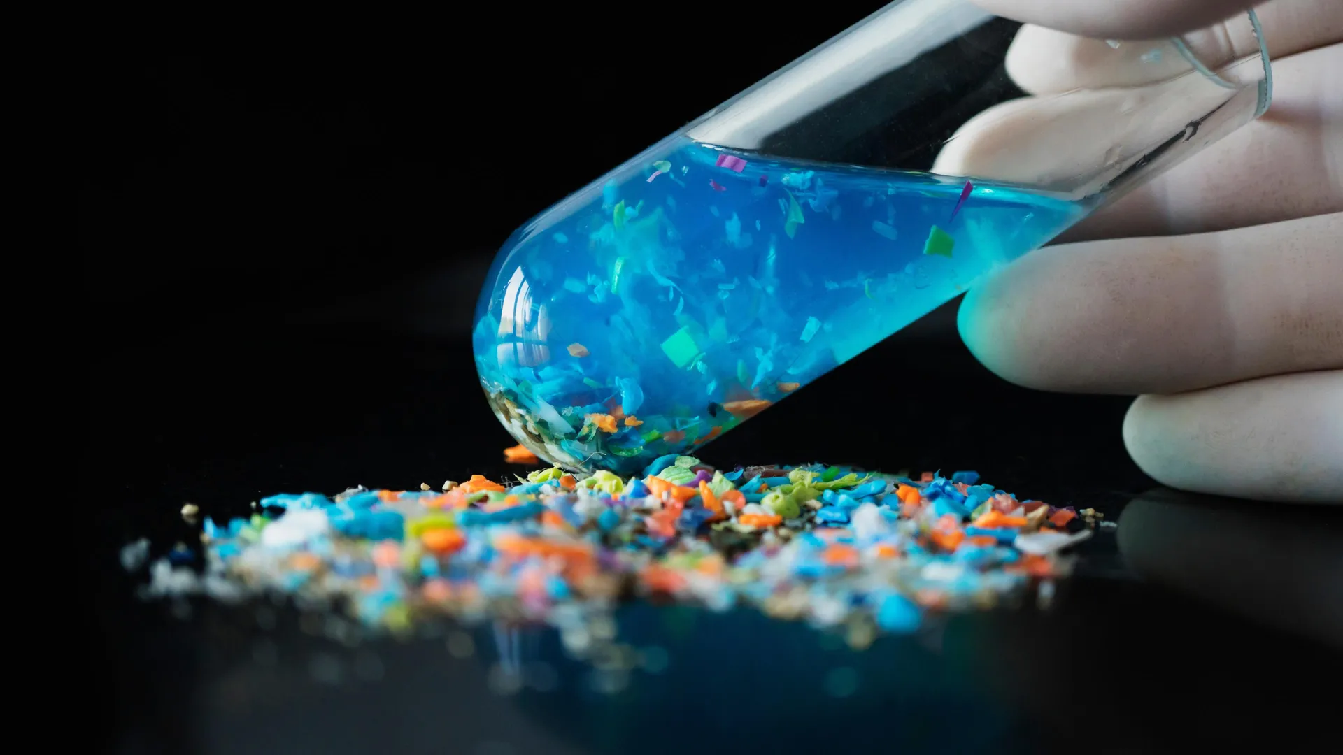
The global production of plastics has surged to unprecedented levels, exceeding 460 million tons annually, a figure that underscores the scale of potential environmental contamination. Each year, millions of tons of these microscopic plastic fragments are released into ecosystems, subsequently finding their way into the digestive tracts and tissues of a vast array of organisms. Scientific investigations have already documented the presence of these particles in diverse marine fauna, avian species, and human tissues, including blood, liver tissue, and even samples from the brain. Preliminary laboratory investigations have suggested potential correlations between microplastic exposure and detrimental health outcomes such as inflammation, organ damage, and developmental abnormalities. Nevertheless, a critical void in scientific understanding persists regarding the precise behavior and impact of these particles once they have entered living organisms.
Existing methodologies for microplastic detection, as explained by researchers, typically provide only a static, or "snapshot," view of the situation. These conventional approaches, which include widely employed techniques like infrared spectroscopy and mass spectrometry, necessitate the destructive processing of biological tissue samples to facilitate analysis. This inherent limitation precludes any direct observation of the dynamic processes involved as particles travel, aggregate, or transform over time within a living organism. While fluorescence imaging has been explored as a potential alternative for real-time tracking, current labeling techniques often suffer from significant drawbacks, including the rapid fading of fluorescent signals, the leakage of dyes from the particles, or a substantial reduction in luminescence intensity when subjected to the complex chemical milieu of biological environments.
In response to these persistent challenges, a dedicated research team has conceptualized and developed a novel approach they term a "fluorescent monomer controlled synthesis strategy." Instead of relying on external fluorescent dyes to coat existing plastic particles, this innovative method involves the direct integration of light-emitting molecular components into the very polymer structure of the plastic during its formation. The core of this technique hinges on the utilization of aggregation-induced emission (AIE) materials, a class of compounds renowned for their remarkable property of exhibiting significantly enhanced fluorescence when their molecules aggregate or cluster together. This intrinsic design feature ensures the generation of a robust and stable fluorescent signal, crucially mitigating the common issue of brightness decay that plagues traditional fluorescence imaging in biological contexts.
This meticulously engineered technique offers researchers an unprecedented degree of control over several key particle characteristics. Scientists can precisely adjust the intensity of the emitted light, select specific wavelengths to determine the color of the fluorescence, and tailor the size and shape of the microplastic particles themselves. A significant advantage of this approach is that the luminescent material is uniformly distributed throughout the entirety of each plastic particle. This uniform distribution ensures that not only intact microplastics but also the progressively smaller fragments generated as they undergo degradation remain continuously visible under the microscope. This capability opens a crucial avenue for tracking the complete lifecycle of microplastics within an organism, from the initial moment of ingestion through their internal journey, subsequent transformations, and eventual breakdown into even smaller constituents.
While this sophisticated strategy is currently undergoing experimental validation, its foundational principles are firmly rooted in established knowledge derived from polymer chemistry and the field of biocompatible fluorescence imaging. The researchers express optimism that this method will emerge as an indispensable instrument for in-depth investigations into the complex interactions between microplastics and cellular structures, tissues, and entire organs.
The precise elucidation of how microplastics are transported and undergo transformation within living organisms is absolutely paramount for accurately assessing their true ecological ramifications and potential risks to human health, according to lead investigators. The ability to dynamically track these particles represents a significant leap forward, enabling a transition from merely quantifying exposure levels to a far more profound comprehension of the underlying mechanisms of toxicity.
As global concerns regarding the escalating problem of plastic pollution continue to intensify, the development and widespread adoption of advanced tools capable of revealing the intricate behavior of microplastics within biological systems are expected to play a pivotal role. Such innovations are anticipated to significantly enhance the accuracy of risk assessments and provide crucial data to inform the development of more effective environmental regulations and mitigation strategies for the future. This novel fluorescence technique offers a beacon of hope in understanding and addressing one of the most pressing environmental and health challenges of our time.