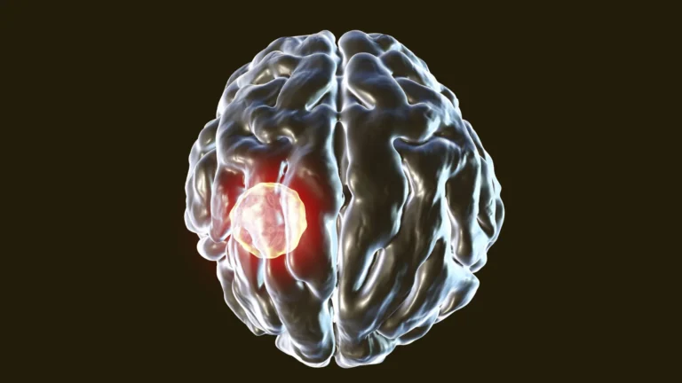
For nearly a decade, the scientific community has been captivated by a revolutionary concept: harnessing the power of bioluminescence to observe the intricate workings of the brain as they unfold. The prevailing paradigm in neuroscience imaging typically involves directing external light sources onto brain tissue to detect cellular activity, often through a process known as fluorescence. This method, while instrumental, presents inherent limitations. As Professor Christopher Moore of Brown University’s Carney Institute for Brain Science eloquently articulated, the fundamental question that sparked this endeavor was, "What if we could illuminate the brain from within?" Traditional fluorescence techniques, while valuable for measuring or stimulating cellular responses, often necessitate sophisticated instrumentation, complex experimental setups, and can yield a lower success rate in capturing fleeting neural events. Recognizing these challenges, researchers envisioned an alternative: enabling neurons themselves to generate light, thereby circumventing the drawbacks of external illumination.
This ambitious vision served as the genesis for the establishment of the Bioluminescence Hub at Brown University’s Carney Institute for Brain Science in 2017. Bolstered by substantial financial backing from the National Science Foundation, this collaborative initiative united leading experts from various institutions. Among the key contributors were Professor Moore, who serves as the associate director of the Carney Institute, Diane Lipscombe, the institute’s director, Ute Hochgeschwender from Central Michigan University, and Nathan Shaner from the University of California San Diego. Their collective mission was to pioneer and disseminate novel neuroscience tools that would imbue nervous system cells with the capacity to both emit light and respond to it, thereby creating an internal signaling system for observation.
A significant milestone in this pursuit was the development of an innovative bioluminescent imaging tool, detailed in a groundbreaking study published in the esteemed journal Nature Methods. This sophisticated instrument, christened the Ca2+ BioLuminescence Activity Monitor, or "CaBLAM," possesses the remarkable ability to capture neural activity at an unprecedented speed, even within individual cells and minuscule cellular compartments. Demonstrating its efficacy in living organisms, CaBLAM has proven successful in mice and zebrafish, enabling continuous recordings that can extend for several hours without the need for any external light input. The design of the core molecular mechanism underpinning CaBLAM was spearheaded by Professor Nathan Shaner, a distinguished neuroscientist and pharmacologist at UC San Diego. Professor Moore lauded CaBLAM as an "amazing molecule" created by Shaner, stating it truly lived up to its ambitious nomenclature.
Understanding the dynamics of living brain cells is paramount to unraveling the complexities of organismal function, a fact underscored by Professor Moore. Current methodologies predominantly rely on genetically encoded calcium-ion indicators that leverage fluorescence. In essence, fluorescence imaging operates by exciting a molecule with an incoming light beam of a specific wavelength, prompting it to emit light at a different wavelength. By engineering these molecules to be sensitive to calcium ions, researchers can detect the presence and concentration of calcium by observing shifts in the emitted light’s intensity or color. This approach, while widely adopted, is not without its significant drawbacks. The prolonged exposure of delicate brain tissue to high-intensity external light can inflict cellular damage. Moreover, the continuous illumination can gradually degrade the fluorescent molecules themselves, a phenomenon known as photobleaching, which diminishes their light-emitting capacity and thereby curtails the duration of experimental observation. Compounding these issues, the delivery of light to the brain necessitates specialized equipment such as lasers and optical fibers, rendering experiments more invasive and technically demanding.
In contrast, bioluminescent imaging offers a fundamentally different and more advantageous approach. Light is intrinsically generated through an enzymatic reaction that breaks down a specific small molecule, thereby obviating the need for external light sources. This internal generation of light eliminates the risks of photobleaching and phototoxic damage, ensuring a far gentler and safer method for studying the fragile architecture of the brain. Furthermore, bioluminescence yields significantly cleaner and more interpretable imaging data. As Professor Shaner explained, brain tissue naturally exhibits a faint intrinsic glow when exposed to external light, which can create background noise that interferes with signal detection. Light also undergoes scattering within brain tissue, blurring both the incoming excitation light and the returning signal, resulting in dimmer, fuzzier images that are particularly challenging to interpret when probing deeper brain structures. Because the brain does not naturally produce bioluminescence, engineered neurons that emit their own light stand out distinctly against a dark background with minimal interference. In essence, bioluminescent neurons act as self-contained light sources, allowing researchers to focus solely on the emitted light, which is far easier to detect and interpret even when it has traversed through scattering tissue. While the scientific community has contemplated the use of bioluminescence for brain activity studies for decades, the crucial challenge of achieving sufficiently bright and sustained light emission for detailed imaging remained unaddressed until this recent breakthrough.
The insights gleaned from this research have been pivotal in making CaBLAM a reality. Professor Moore expressed his enthusiasm for the current findings, emphasizing that these novel molecular constructs have, for the first time, granted researchers the ability to observe the activation of individual cells in isolation, akin to using an exceptionally sensitive and specialized movie camera to record neural activity in real-time. With CaBLAM, scientists can now meticulously document the behavior of a single neuron within a living organism, including the dynamic activity occurring within its distinct cellular components. The study reported an unprecedented continuous recording session lasting five hours, a feat that would be entirely unattainable with conventional fluorescence-based techniques. Professor Moore highlighted that bioluminescence significantly streamlines the study of complex behaviors and learning processes by enabling the capture of entire phenomena with a substantially reduced reliance on external hardware.
The CaBLAM project represents a broader strategic objective within the Bioluminescence Hub: to develop innovative methods for both observing and actively controlling neural activity. One intriguing line of research involves engineering living cells to emit light signals that can be detected by neighboring cells, thereby enabling neurons to communicate through light itself – a concept Professor Moore playfully terms "rewiring the brain with light." The team is also advancing techniques that utilize calcium ions to modulate cellular activity. As these diverse research threads have progressed, a common prerequisite emerged: the necessity for more potent and efficient calcium sensors. This critical requirement has become a central tenet of the hub’s ongoing research endeavors. Professor Moore emphasized the hub’s commitment to serving as a catalyst for progress in the field by ensuring the development of these foundational technological components.
Professor Moore anticipates that the applications of CaBLAM will extend far beyond the realm of neuroscience, offering new avenues for investigating activity in other physiological systems throughout the body. He believes this technological advancement unlocks a "whole new range of options for seeing how the brain and body work," including the capacity to simultaneously monitor activity across multiple bodily regions. This significant achievement also underscores the profound impact of collaborative scientific inquiry. The project benefited from the contributions of at least 34 scientists, representing a consortium of Bioluminescence Hub partners such as Brown University, Central Michigan University, UC San Diego, the University of California Los Angeles, and New York University. The research was made possible through vital funding from esteemed organizations including the National Institutes of Health, the National Science Foundation, and the Paul G. Allen Family Foundation.