For decades, the intricate network of human blood vessels, essential conduits for life-sustaining blood flow, presented a significant modeling challenge in scientific research. While these biological structures exhibit remarkable adaptability—bending, branching, narrowing, and widening in a dynamic fashion—conventional laboratory representations often simplified them into static, uniform tubes. This fundamental disparity between simplified models and physiological reality created a persistent gap in understanding the genesis and progression of many vascular diseases, which frequently originate and thrive in areas characterized by complex blood flow patterns and unique architectural features. Recognizing this critical limitation, a team of researchers within the Department of Biomedical Engineering at Texas A&M University has unveiled a groundbreaking customizable vessel-chip system designed to bridge this gap, offering an unprecedented platform for studying cardiovascular pathologies and accelerating the development of novel therapeutic interventions.
The human circulatory system is a masterpiece of biological engineering, comprising arteries, veins, and capillaries that collectively stretch over 60,000 miles within an adult body. Its complexity extends beyond mere length, encompassing a myriad of geometric variations—from sharp bifurcations where vessels split, to gradual curves, and localized anomalies such as sudden expansions (aneurysms) or constrictions (stenoses). Each of these architectural features profoundly influences hemodynamics, the study of blood flow, which in turn dictates the mechanical forces exerted on the vessel walls, particularly shear stress. Endothelial cells, the specialized cells lining the inner surface of blood vessels, are exquisitely sensitive to these mechanical cues. Abnormal or disturbed shear stress, often found at branches, curves, and regions of altered vessel diameter, is a known precursor and driver of numerous vascular diseases, including atherosclerosis, the hardening and narrowing of arteries, and the formation of potentially fatal aneurysms. Traditional straight-tube models, by their very nature, failed to adequately replicate these critical flow disturbances, thus limiting their utility in deciphering disease mechanisms in their natural, complex context.
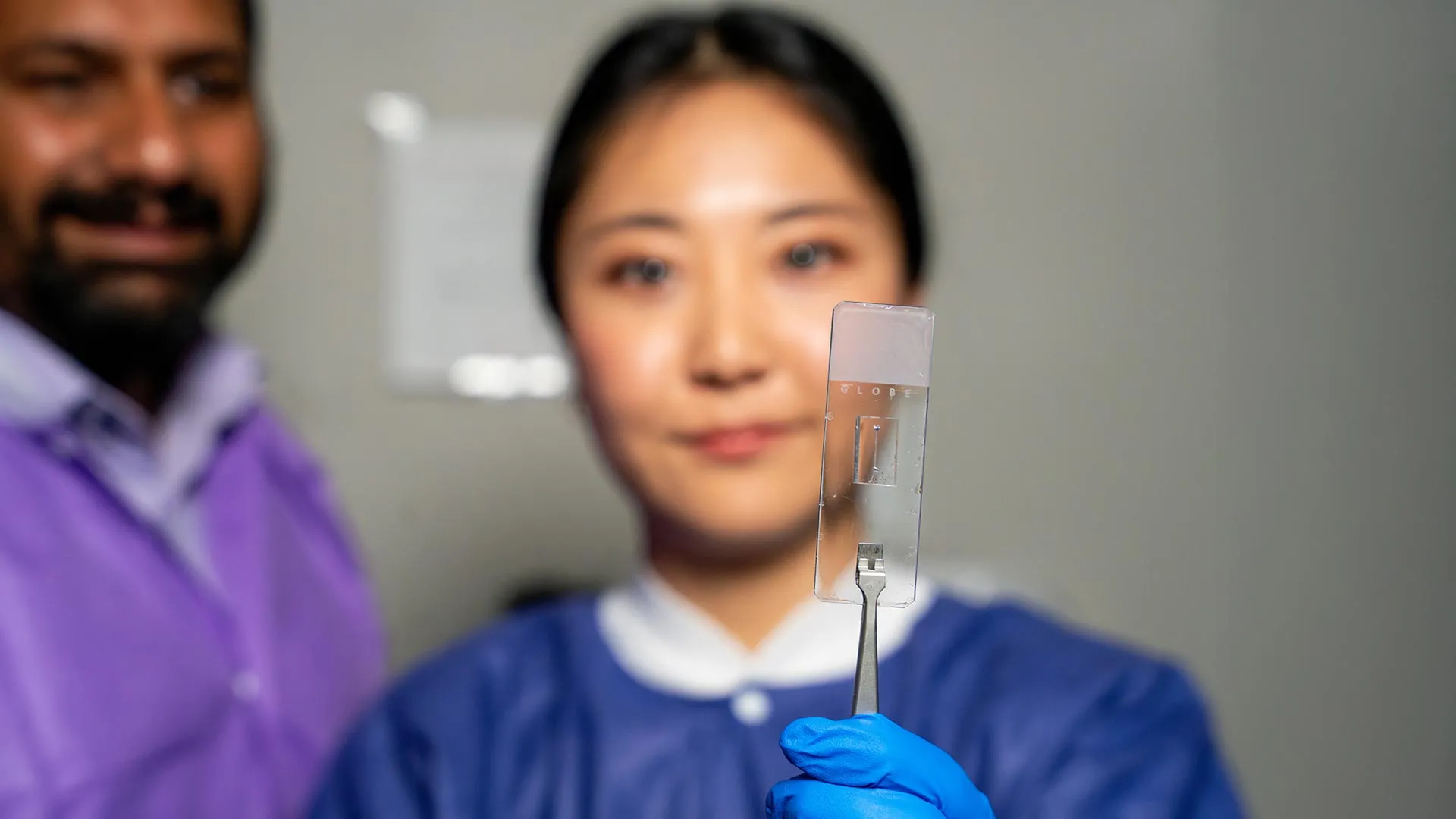
The innovative work spearheaded by Dr. Abhishek Jain, an associate professor and the Barbara and Ralph Cox ’53 faculty fellow in biomedical engineering, alongside his dedicated team in the Bioinspired Translational Microsystems Laboratory, directly addresses this long-standing challenge. Their novel vessel-chip system is a sophisticated microfluidic device engineered to faithfully mimic the microarchitecture and dynamic environment of human blood vessels on a vastly reduced scale. Unlike its predecessors, this advanced iteration is not confined to linear geometries. It empowers scientists to fabricate complex, three-dimensional vascular structures that precisely mirror the anatomical irregularities where vascular diseases are most likely to manifest. This capability represents a significant leap forward, providing a more physiologically relevant setting for investigating disease initiation and progression.
At the heart of this technological breakthrough is the meticulous design work contributed by Jennifer Lee, a master’s student in biomedical engineering. Building upon earlier foundational research within the same laboratory, which included the development of a straight vessel-chip design by former graduate student Dr. Tanmay Mathur, Lee’s contribution significantly expands the repertoire of reproducible vascular geometries. "We wanted to model the diverse types of vessels that exist in the human body," Lee explained, highlighting the critical importance of these variations. "There are branched vessels, or aneurysms that have sudden expansion, and then stenosis that restricts the vessel. All these different types of vessels cause the blood flow pattern to be significantly changed, and the inside of the blood vessel is affected by the level of shear stress caused by these flow patterns." By precisely replicating these conditions, researchers can now observe cellular responses and pathological changes in an environment that closely mirrors the human body, a capability previously unattainable with simplified models.
The "organ-on-a-chip" paradigm, a rapidly evolving field of biomedical engineering, underpins this advancement. These microfluidic devices integrate living cells within micro-engineered environments to recapitulate the physiological functions and mechanical properties of entire organs or organ systems. The vessel-chip system exemplifies the pinnacle of this technology, offering several compelling advantages. Firstly, it provides a non-animal platform for research, addressing ethical concerns associated with animal testing while potentially yielding more human-relevant data. Secondly, its customizability opens avenues for personalized medicine, allowing researchers to create patient-specific vascular models using an individual’s own cells, thereby predicting drug responses or disease trajectories with greater accuracy. This precision is particularly crucial in complex conditions like atherosclerosis, where individual genetic predispositions and lifestyle factors interact with local hemodynamic forces to drive disease.
The implications for drug discovery and development are profound. Pharmaceutical companies and academic researchers often struggle with the translational gap—findings from preclinical animal studies frequently fail to translate into effective human therapies. The Texas A&M vessel-chip, with its ability to incorporate actual cellular and tissue material, offers a more predictive in vitro model. New therapeutic agents, whether they are designed to reduce inflammation, prevent clot formation, or enhance vessel repair, can now be tested in complex vascular environments that more closely simulate the disease state in humans. This could significantly de-risk drug development, reduce costs, and accelerate the pipeline for innovative treatments for cardiovascular diseases, which remain the leading cause of mortality worldwide.
The project’s significance is further underscored by its publication in the prestigious journal Lab on a Chip, where it is slated to appear on the cover of the May 2025 issue. This recognition within the scientific community highlights the innovative nature and potential impact of the research. Dr. Jain emphasized the transformative potential, stating, "We can now start learning about vascular disease in ways we’ve never been able to before. Not only can you make these structures complex, you can put actual cellular and tissue material inside them and make them living. These are the sites where vascular diseases tend to develop, so understanding them is critical." This ability to create "living" complex structures, complete with endothelial cells, allows for real-time observation of cellular interactions, gene expression changes, and the development of pathological hallmarks under controlled, yet physiologically relevant, conditions.
Looking ahead, the research team is not content to rest on its laurels. While the current model primarily incorporates endothelial cells, future iterations are envisioned to integrate additional cell types critical to vascular biology, such as smooth muscle cells, fibroblasts, and even immune cells. This expansion would allow for a more holistic understanding of how different tissue components interact within the vascular wall and with the flowing blood, replicating the complex cellular ecosystem of real blood vessels. Dr. Jain refers to this ambitious future direction as "the fourth dimensionality of organs-on-a-chip," a concept that extends beyond merely replicating cells and flow to encompass the dynamic interaction of cells and flow within increasingly complex architectural states over time. This holistic approach promises to unlock deeper insights into multifactorial diseases and the intricate regenerative processes of the vasculature.
Beyond the scientific breakthroughs, this project also serves as a testament to the power of mentorship and the cultivation of emerging scientific talent. Jennifer Lee’s journey from an undergraduate honors student seeking hands-on research experience to a published master’s student exemplifies the enriching environment fostered at Texas A&M. Initially unfamiliar with organs-on-a-chip technology, Lee’s curiosity and dedication blossomed under Dr. Jain’s guidance. Her participation in the Master of Science fast-track program provided the framework to transition her initial interest into high-impact, publishable research. Dr. Jain lauded Lee’s commitment, noting, "Jennifer demonstrated perseverance, curiosity, and creativity and started taking up research projects very quickly. Our fast-track program enables students like Jennifer to take on sort of high-impact, high-risk research and not just do a science project, but take it all the way to its outcome and get it published."
The skills honed in such a dynamic research setting extend far beyond technical proficiency. Lee herself attested to the invaluable experience gained in collaboration, communication, and problem-solving. Working alongside peers, graduate students, and postdoctoral researchers within the Bioinspired Translational Microsystems Laboratory provided a crucible for developing essential professional attributes. "It’s such a good environment to interact with not only peers but also graduate students and postdoctoral researchers," she reflected. "You’re able to learn teamwork and communication, work ethic, and just trying different things out. I think it’s such a valuable experience that students have available. We have such good faculty research labs." These interpersonal and critical thinking skills are as crucial to scientific advancement as the technical expertise itself, preparing the next generation of researchers to tackle complex global health challenges.
The broad utility and strategic importance of this research are further evidenced by the diverse array of prestigious organizations providing financial support. Funding has been secured from major entities including the U.S. Army Medical Research Program, NASA, the Biomedical Advanced Research and Development Authority (BARDA), the National Institutes of Health (NIH), the U.S. Food and Drug Administration (FDA), the National Science Foundation (NSF), and the Texas A&M University Office of Innovation Translational Investment Funds. This multi-agency backing underscores the wide-ranging potential applications of the vessel-chip technology, from understanding vascular responses in extreme environments like space to developing countermeasures for battlefield trauma, enhancing drug safety evaluations, and advancing fundamental biomedical knowledge.
In conclusion, the development of this customizable, living vessel-chip system by the Texas A&M team represents a pivotal moment in vascular research. By moving beyond simplistic models to embrace the true anatomical and hemodynamic complexity of human blood vessels, scientists are now equipped with an unprecedented tool to unravel the mysteries of cardiovascular diseases. This innovative platform promises to accelerate drug discovery, facilitate the realization of personalized medicine, and ultimately improve patient outcomes, heralding a new era of precision in understanding and treating the body’s vital circulatory network.