
A groundbreaking advancement in neuroscience has emerged with the development of a sophisticated protein capable of capturing the subtle chemical conversations that brain cells receive, a feat previously eluding scientific observation. This novel protein offers a critical new lens into brain function, moving beyond the examination of outgoing signals to meticulously record the incoming chemical messages that neurons process. These vital incoming signals are primarily orchestrated by glutamate, a paramount neurotransmitter indispensable for the intricate tapestry of brain communication. While glutamate’s role in fundamental cognitive processes such as learning and memory is well-established, its fleeting and exceedingly faint nature has rendered its direct measurement an immense challenge for researchers.
The introduction of this innovative protein sensor fundamentally alters the landscape of neuroscientific inquiry, empowering scientists to detect and quantify these subtle chemical influxes as they occur. This capability grants unprecedented access to a crucial dimension of neural interaction that has, until now, remained largely obscured. The ability to directly observe the reception of signals allows for a profound exploration into the intricate mechanisms by which individual neurons process incoming information. Each neuron in the brain is bombarded by thousands of distinct inputs, and the manner in which it integrates these diverse signals ultimately dictates its own subsequent output. This complex computational process is believed to be the very foundation of our thoughts, decisions, and memories, and its direct study promises to unravel how the brain executes its most sophisticated operations.
Beyond its implications for understanding normal brain function, this breakthrough holds immense promise for advancing research into neurological and psychiatric disorders. Dysregulation of glutamate signaling has been implicated in a spectrum of debilitating conditions, including Alzheimer’s disease, schizophrenia, autism spectrum disorder, and epilepsy. By enabling more precise measurement of these signals, researchers are poised to pinpoint the underlying biological anomalies that contribute to the pathogenesis of these disorders, potentially paving the way for novel diagnostic and therapeutic strategies. Furthermore, the pharmaceutical industry stands to benefit significantly from this development. Experimental treatments designed to modulate neural activity can now be rigorously evaluated for their precise impact on real-time synaptic function, accelerating the discovery and refinement of more effective therapeutic interventions.
The engineered protein, christened iGluSnFR4 (pronounced "glue sniffer"), is the product of a collaborative effort between researchers at the Allen Institute and HHMI’s Janelia Research Campus. Functioning as a highly sensitive molecular indicator for glutamate, iGluSnFR4 possesses the remarkable capacity to detect even the faintest incoming signals exchanged between neurons. This unparalleled sensitivity allows scientists to meticulously chart the release and reception of glutamate, providing a novel framework for interpreting the complex patterns of neural activity that underpin learning, memory formation, and emotional processing. The ability to visualize neuronal communication within the living brain in real time marks a paradigm shift in neuroscience. These findings, recently disseminated in the esteemed journal Nature Methods, are anticipated to profoundly reshape the methodologies employed in measuring and analyzing neural activity.
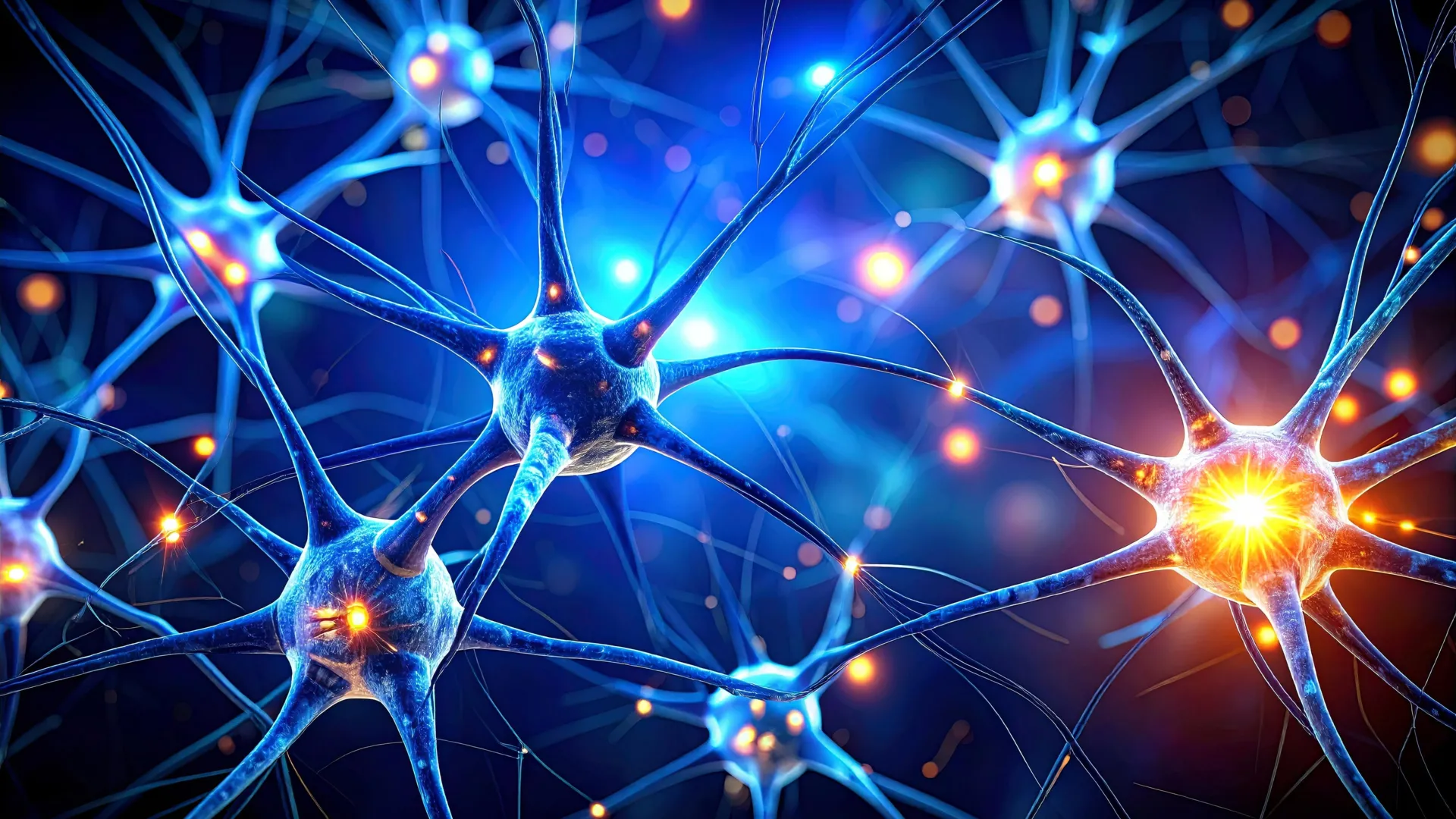
To fully appreciate the significance of this scientific leap, it is essential to grasp the fundamental principles of neuronal communication. The human brain comprises billions of neurons, interconnected cells that transmit information through electrical impulses propagated along specialized extensions known as axons. When an electrical signal traverses an axon and reaches its terminus, it encounters a minuscule gap called a synapse, separating it from the next neuron. This electrical signal cannot directly bridge this gap.
Instead, the arrival of the electrical impulse at the axon terminal triggers the release of neurotransmitters, chemical messengers, into the synaptic cleft. Among these messengers, glutamate stands out as the most prevalent and arguably the most critical. Its pivotal role in synaptic plasticity, the cellular basis of learning and memory, and its involvement in a vast array of cognitive functions, including emotion and perception, are well-documented. Upon diffusing across the synapse and binding to receptors on the postsynaptic neuron, glutamate can excite that neuron, prompting it to generate its own electrical signal and propagate the communication chain.
This intricate process can be likened to a cascade of falling dominos, albeit one of vastly greater complexity and nuance. Each neuron is a recipient of input from thousands of other neurons, and its decision to fire an action potential is contingent upon the integration of a specific constellation of incoming signals. The development of iGluSnFR4 now empowers scientists to directly identify which patterns of incoming glutamatergic activity are responsible for triggering a particular postsynaptic response.
Historically, observing these incoming signals within living brain tissue presented a formidable obstacle. Existing technologies, while valuable, often suffered from limitations in speed or sensitivity, proving inadequate for capturing the rapid and localized events occurring at individual synapses. Consequently, researchers were largely confined to observing fragmented glimpses of the communication process, rather than the complete, dynamic exchange between neurons. The advent of iGluSnFR4 fundamentally overcomes this limitation, offering the unprecedented ability to witness the entirety of the neural conversation as it unfolds.
Dr. Kaspar Podgorski, a leading author of the study and a senior scientist at the Allen Institute, eloquently articulated the transformative impact of this technology, comparing the pre-iGluSnFR4 era to attempting to comprehend a book with all its words jumbled and its grammatical structure obliterated. "I feel like what we’re doing here is adding the connections between those neurons and by doing that, we now understand the order of the words on the pages, and what they mean," he explained. Prior to the availability of protein sensors like iGluSnFR4, neuroscientists possessed sophisticated methods for assessing the structural connectivity between neurons, and in separate experimental paradigms, could measure the outgoing signals emitted by some neurons. However, integrating these two distinct streams of information – the physical connections and the outgoing transmissions – remained a significant challenge. The capacity to measure what neurons are "saying" to specific other neurons was lacking. "What we have invented here is a way of measuring information that comes into neurons from different sources, and that’s been a critical part missing from neuroscience research," Dr. Podgorski emphasized.
The triumph of iGluSnFR4 is a testament to the power of interdisciplinary collaboration. Dr. Jeremy Hasseman, a scientist at HHMI’s Janelia Research Campus, highlighted the synergistic relationship that fueled this breakthrough: "The success of iGluSnFR4 stems from our close collaboration started at HHMI’s Janelia Research Campus between the GENIE Project team and Kaspar’s lab. That research has extended to the phenomenal in vivo characterization work done by the Allen Institute’s Neural Dynamics group. This was a great example of collaboration across labs and institutes to enable new discoveries in neuroscience."
This scientific leap effectively bridges a critical chasm in modern neuroscience, rendering direct observation of how neurons assimilate information an achievable reality. With iGluSnFR4 now readily accessible to the scientific community through Addgene, researchers are equipped with an exceptionally potent instrument for dissecting the complexities of brain function with unparalleled detail. As this technology proliferates and is integrated into a wider array of research endeavors, it is poised to illuminate answers to some of neuroscience’s most enduring and profound questions, ushering in a new era of understanding the human brain.


