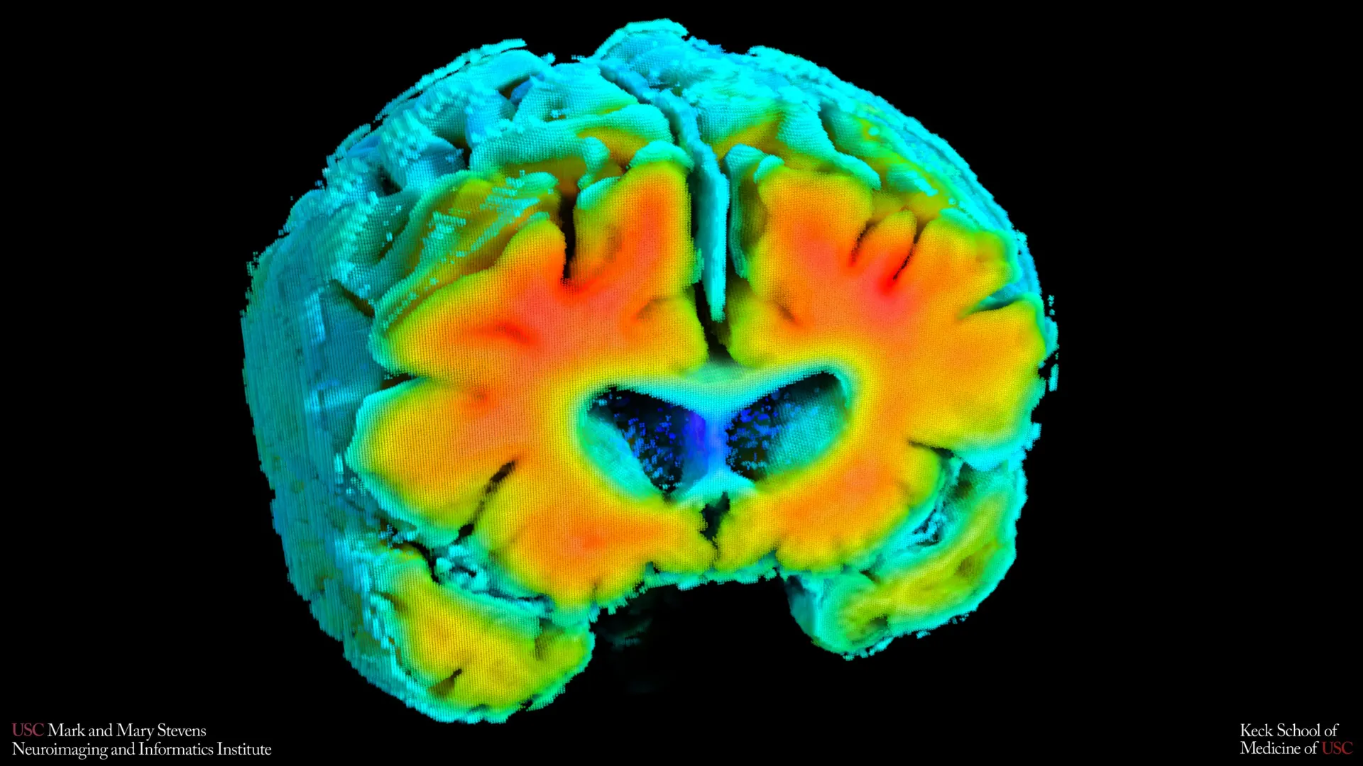
A groundbreaking investigation conducted by researchers at the Mark and Mary Stevens Neuroimaging and Informatics Institute (Stevens INI), part of the Keck School of Medicine of USC, suggests a profound and often overlooked connection between the subtle dynamics of cerebral blood flow and the earliest stages of Alzheimer’s disease. The study’s findings illuminate how minute alterations in the brain’s vascular system, specifically concerning blood circulation and oxygen delivery to neural tissues, may precede the more commonly recognized neuropathological markers of Alzheimer’s, thereby offering a novel perspective on disease initiation and progression. This research, recently featured in the esteemed journal Alzheimer’s & Dementia: The Journal of the Alzheimer’s Association, delves into the intricate relationship between vascular integrity and the neurodegenerative cascade, proposing that compromised cerebrovascular function could serve as a critical, early indicator of risk.
The brain, a metabolically demanding organ, relies unequivocally on a constant and robust supply of oxygen and nutrients delivered via its extensive vascular network. Any disruption to this delicate system can have far-reaching consequences, potentially initiating or accelerating neurodegenerative processes. For years, the scientific community has largely focused on the accumulation of amyloid-beta plaques and tau neurofibrillary tangles as the primary drivers of Alzheimer’s pathology. However, the current study underscores a compelling argument for integrating cerebrovascular health into the foundational understanding of the disease, suggesting that impairments in how blood traverses the brain and how oxygen is effectively utilized by brain cells may represent an initial, ‘silent’ phase of the illness, long before cognitive symptoms manifest.
Investigators meticulously examined a cohort of older adults, encompassing individuals with intact cognitive function as well as those presenting with varying degrees of cognitive impairment. Their innovative approach utilized simple, non-invasive diagnostic modalities to quantify key aspects of cerebral blood flow and tissue oxygenation. The data revealed compelling associations between these cerebrovascular metrics and established neuropathological hallmarks of Alzheimer’s disease. Specifically, individuals exhibiting healthier patterns of brain blood flow and oxygen metabolism tended to have lower levels of amyloid plaque deposition within the brain parenchyma and maintained larger hippocampal volumes—both factors independently linked to a reduced propensity for Alzheimer’s. The hippocampus, a bilateral structure nestled deep within the medial temporal lobe, is indispensable for memory consolidation and spatial navigation, and its atrophy is a well-documented feature of Alzheimer’s pathology, correlating strongly with cognitive decline.
Amaryllis A. Tsiknia, the lead author of this pivotal study and a USC PhD candidate, emphasized the significance of these vascular contributions. "While amyloid and tau proteins have historically been at the forefront of Alzheimer’s research as primary pathological agents, our findings unequivocally demonstrate the critical role played by cerebral blood flow and oxygen supply," Tsiknia stated. "The data indicates that when the brain’s intricate vascular system functions optimally, mirroring patterns observed in healthy aging, we concomitantly observe brain characteristics that are intrinsically linked to superior cognitive health and resilience against neurodegeneration." This statement highlights a paradigm shift, urging a more holistic view of Alzheimer’s etiology that extends beyond proteinopathies to encompass systemic physiological factors.
To achieve these insights, the research team pioneered the application of two sophisticated yet entirely non-invasive techniques. The first, Transcranial Doppler (TCD) ultrasonography, is a highly effective method for continuously monitoring the velocity of blood flow through the major cerebral arteries. This technique involves placing a small probe on the scalp, which emits and receives ultrasonic waves, allowing real-time assessment of blood dynamics without discomfort or radiation exposure. The second technique, Near-Infrared Spectroscopy (NIRS), offers a complementary measure by evaluating the efficiency of oxygen delivery and utilization within superficial layers of the cerebral cortex. NIRS employs infrared light to assess changes in the concentration of oxygenated and deoxygenated hemoglobin, providing a window into regional brain oxygenation. Both procedures are conducted while the participant is at rest, requiring minimal cooperation and making them highly amenable to widespread clinical application.
Subsequent to data acquisition, the research team employed advanced mathematical modeling to synthesize the readings from TCD and NIRS into comprehensive indicators of cerebrovascular function. These sophisticated algorithms allowed them to derive metrics that accurately reflect the brain’s adaptive capacity—its ability to finely tune blood flow and oxygen provision in dynamic response to natural physiological fluctuations in systemic blood pressure and carbon dioxide levels. This adaptive capacity is a hallmark of a healthy vascular system, ensuring metabolic demands are met efficiently across diverse physiological states.
The study’s robust findings further elucidated that participants whose vascular indicators closely aligned with those of cognitively healthy older adults consistently presented with lower cerebral amyloid burden and preserved hippocampal volume. These structural and biochemical features are widely recognized as critical markers associated with a substantially reduced risk of developing Alzheimer’s disease. Meredith N. Braskie, PhD, senior author of the study and an assistant professor of neurology at the Keck School of Medicine, underscored the clinical relevance of these findings. "These novel vascular measures are capturing profoundly meaningful information regarding overall brain health," Braskie remarked. "They exhibit a remarkable concordance with observations derived from more established, albeit more resource-intensive, imaging modalities such such as Magnetic Resonance Imaging (MRI) and Positron Emission Tomography (PET) scans, which are standard tools in Alzheimer’s research. This provides crucial evidence regarding the intricate interrelationship between cerebrovascular health and the conventional neuropathological markers of Alzheimer’s disease risk."
Furthermore, the investigators observed a clear gradient: individuals formally diagnosed with mild cognitive impairment (MCI) or clinically manifest dementia consistently demonstrated measurably weaker cerebrovascular function compared to their cognitively normal counterparts. This observation lends substantial support to the hypothesis that a decline in the health and efficiency of the brain’s blood vessels is not merely a consequence but an integral component of the broader Alzheimer’s disease continuum, potentially preceding and contributing to the overt neurodegenerative changes.
Dr. Arthur W. Toga, PhD, director of the Stevens INI, highlighted the broader implications of these discoveries. "These findings significantly augment the burgeoning body of evidence indicating that Alzheimer’s disease pathology encompasses meaningful vascular contributions, operating in concert with, or perhaps even preceding, the classic neurodegenerative changes attributed to amyloid and tau," Toga asserted. "Developing a comprehensive understanding of how the regulation of cerebral blood flow and oxygen interacts with amyloid pathophysiology and brain structural integrity opens entirely new avenues for both the early detection of Alzheimer’s risk and, crucially, for the development of innovative preventative strategies."
One of the most compelling aspects of this research lies in the potential for earlier and more widespread screening for Alzheimer’s risk. Unlike the current gold-standard diagnostic tools—such as PET imaging, which involves radioactive tracers and high costs, or MRI, which can be time-consuming and inaccessible to all patients—the Transcranial Doppler ultrasound and Near-Infrared Spectroscopy methods are considerably less expensive, operationally simpler, and can be performed in diverse clinical settings. They do not necessitate intravenous injections, expose patients to ionizing radiation, or require patients to perform demanding cognitive tasks. This inherent simplicity and safety profile makes these non-invasive techniques particularly well-suited for large-scale population screening initiatives, or for individuals who may be unable to undergo more intensive or restrictive brain imaging procedures due to physical limitations, claustrophobia, or financial constraints.
It is important to note, as the authors prudently caution, that the current findings represent a singular cross-sectional snapshot in time. As such, while they establish strong correlations, they do not definitively prove a direct cause-and-effect relationship. To address this, ongoing longitudinal studies are actively tracking participants over extended periods. The primary objective of these follow-up investigations is to determine whether subtle shifts or deteriorations in these novel vascular measures can reliably predict future cognitive decline or assess an individual’s response to emerging therapeutic interventions aimed at slowing or halting the progression of Alzheimer’s.
Tsiknia articulated the future vision for this research: "If we can effectively monitor these specific vascular signals over time, we may gain the unprecedented ability to identify individuals at a significantly elevated risk for Alzheimer’s at a much earlier stage. This foundational insight would then allow us to rigorously test whether targeted interventions designed to improve or sustain optimal vascular health can effectively mitigate or even reverse Alzheimer’s-related brain changes, potentially altering the disease trajectory for millions." This forward-looking perspective positions cerebrovascular health not just as a diagnostic biomarker, but as a potential therapeutic target, opening new frontiers in the fight against Alzheimer’s disease.
The growing understanding of the neurovascular unit—the complex interplay between neurons, astrocytes, and the intricate network of blood vessels that supply them—is revolutionizing neuroscience. This study adds significant weight to the argument that disruptions within this unit are not merely collateral damage in neurodegenerative diseases but may, in fact, be instrumental in their genesis and propagation. By focusing on the health of the brain’s vasculature, researchers are paving the way for a more comprehensive and proactive approach to managing Alzheimer’s disease, shifting the focus from treating symptoms to preventing the disease before it takes hold.
This vital research was significantly supported by funding from the Office of The Director, National Institutes of Health, under Award Number S10OD032285, and by the National Institute on Aging under Award Number R01AG058162. In addition to Amaryllis A. Tsiknia and Meredith N. Braskie, the collaborative efforts of Peter S. Conti, Rebecca J. Lepping, Brendan J. Kelley, Rong Zhang, Sandra A. Billinger, Helena C. Chui, and Vasilis Z. Marmarelis were instrumental in the successful execution and interpretation of this study. Their combined expertise has propelled our understanding of Alzheimer’s disease into a new, vascular-centric era.