
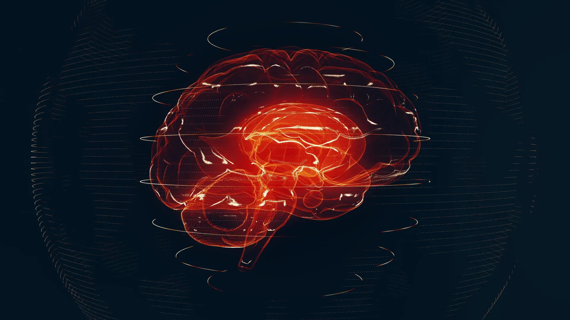
A groundbreaking artificial intelligence system developed at the University of Michigan is poised to redefine the landscape of neurological diagnostics, capable of scrutinizing brain magnetic resonance imaging (MRI) scans with unprecedented speed and accuracy. This novel technology, detailed in a recent publication in Nature Biomedical Engineering, has demonstrated the ability to identify a wide spectrum of neurological conditions with a diagnostic precision reaching an impressive 97.5%. Beyond mere identification, the system also possesses the crucial capability to ascertain the urgency of medical intervention required by patients, a critical factor in optimizing treatment pathways for time-sensitive conditions.
The implications of this innovation are far-reaching, with researchers suggesting it could fundamentally alter how brain imaging is managed within healthcare infrastructures across the United States. The escalating global demand for MRI services, coupled with the considerable strain it places on both physicians and healthcare systems, creates a compelling need for advanced solutions. This AI model, christened "Prima," offers a potent remedy by promising to alleviate diagnostic burdens and enhance treatment efficacy through the provision of rapid and precise information. Dr. Todd Hollon, a senior author of the study and a neurosurgeon at University of Michigan Health, highlighted the potential for Prima to streamline workflows and improve patient outcomes, particularly in high-volume or resource-limited settings.
The development and rigorous testing of Prima involved a comprehensive evaluation over a one-year period, during which the system analyzed a vast repository of over 30,000 MRI studies. This extensive dataset allowed researchers to assess Prima’s performance across more than 50 distinct radiologic diagnoses pertaining to major neurological disorders. In head-to-head comparisons with other advanced AI models, Prima consistently delivered superior diagnostic performance. Crucially, its utility extends beyond simply diagnosing diseases; it can effectively prioritize cases, flagging those requiring immediate medical attention.
Conditions such as strokes and brain hemorrhages necessitate swift medical intervention to mitigate potential long-term damage. Prima’s design incorporates an automatic alert system, capable of instantaneously notifying healthcare providers when such critical conditions are detected. This immediate notification mechanism ensures that prompt action can be initiated, potentially saving valuable time and improving patient prognoses. The system is engineered to direct these alerts to the most appropriate subspecialist, whether a stroke neurologist or a neurosurgeon, thereby ensuring that the right expertise is engaged without delay. Patient feedback and diagnostic insights become available immediately following the completion of the imaging procedure, a significant departure from traditional turnaround times.
The paramount importance of accuracy in brain MRI interpretation is undeniable, but researchers underscore that speed is equally vital for timely diagnosis and ultimately, for achieving better patient outcomes. Yiwei Lyu, M.S., a co-first author and postdoctoral fellow in Computer Science and Engineering at the University of Michigan, emphasized that Prima’s results demonstrate its capacity to enhance clinical workflows and expedite care without compromising diagnostic fidelity. This balance between speed and accuracy is a key differentiator for the Prima system.
Prima distinguishes itself as a Vision Language Model (VLM), a sophisticated category of artificial intelligence that possesses the unique ability to process and integrate visual data, such as images and videos, with textual information in real-time. While AI has been applied to MRI analysis previously, Prima represents a departure in its methodological approach. Earlier iterations of AI in this field were often trained on carefully curated, narrow subsets of MRI data, designed to perform highly specific tasks, such as detecting lesions or estimating the risk of dementia. In contrast, Prima was trained on an exceptionally broad and diverse dataset.
The research team at the University of Michigan employed a comprehensive strategy, utilizing virtually every available MRI scan collected since the digitization of radiology records at the institution. This colossal dataset comprised over 200,000 MRI studies and an astonishing 5.6 million imaging sequences. Furthermore, Prima’s training regimen incorporated crucial contextual information, including patients’ clinical histories and the specific reasons documented by physicians for ordering each imaging study. This holistic approach allows Prima to function in a manner analogous to a seasoned radiologist, integrating both imaging findings and patient context to formulate a comprehensive understanding of their health status. Samir Harake, a data scientist and co-first author from Dr. Hollon’s Machine Learning in Neurosurgery Lab, explained that this integrated approach facilitates superior performance across a wide array of predictive tasks.
The development of Prima is particularly timely given the persistent challenges of MRI delays and shortages within the radiology field. Millions of MRI scans are performed annually worldwide, with a substantial proportion dedicated to the investigation of neurological diseases. The demand for these scans is escalating at a rate that significantly outpaces the availability of specialized neuroradiology services. This imbalance has unfortunately led to a cascade of issues, including critical staffing shortages, diagnostic delays that can impact patient care, and an increased risk of errors. In some cases, patients may experience waiting periods of several days, or even longer, for their MRI results to be returned, depending on the healthcare facility.
Dr. Vikas Gulani, co-author and chair of the Department of Radiology at U-M Health, emphasized the urgent need for innovative technological solutions to address these disparities. He noted that whether patients are seeking care at large health systems grappling with overwhelming patient volumes or at rural hospitals with limited resources, advancements are essential to improve access to radiology services. The collaborative efforts of the University of Michigan teams have yielded a cutting-edge solution with the potential for significant scalability and widespread impact.
While Prima has demonstrated remarkable performance in its initial evaluations, researchers are quick to emphasize that this work is still in its early stages of development. Future research will focus on refining the system by incorporating even more granular patient information and data extracted from electronic medical records. This continuous refinement aims to further enhance diagnostic accuracy and broaden the system’s clinical utility. This iterative approach mirrors the complex interpretative processes undertaken by human radiologists and physicians when analyzing MRIs and other medical imaging studies in real-world clinical scenarios.
The integration of AI into healthcare is an ongoing trend, yet many existing systems are confined to performing narrowly defined, single-purpose tasks. Dr. Hollon drew an analogy, describing Prima as akin to "ChatGPT for medical imaging," suggesting that the underlying technological principles could eventually be adapted for a variety of other imaging modalities. This could include applications for mammograms, chest X-rays, and ultrasounds, potentially democratizing advanced diagnostic capabilities across different medical specialties.
The vision for Prima extends beyond that of a standalone diagnostic tool; it is conceptualized as a "co-pilot" for medical imaging interpretation. Similar to how AI assistants can help draft emails or provide recommendations, Prima aims to augment the capabilities of clinicians, providing them with rapid, accurate, and comprehensive insights derived from imaging data. Dr. Hollon articulated his belief that Prima represents a compelling example of the transformative potential inherent in the synergistic integration of healthcare systems and AI-driven models, ultimately driving innovation and improving the quality of health care delivery. The research and development of Prima were supported by a consortium of prestigious funding bodies, including the National Institute of Neurological Disorders and Stroke (part of the National Institutes of Health), the Chan Zuckerberg Initiative (CZI), the Frankel Institute for Heart and Brain Health, the Mark Trauner Brain Research Fund, the Zenkel Family Foundation, Ian’s Friends Foundation, and the UM Precision Health Investigators Awards grant program. It is important to note that the content of this publication represents the views of the researchers and does not necessarily reflect the official positions of the NIH.


