The period of adolescence represents a profound phase of transformation, extending far beyond mere physical and social maturation; it is a critical window for the intricate sculpting of the brain’s neural architecture. During these formative years, sophisticated cognitive functions such as strategic planning, abstract reasoning, and complex decision-making undergo significant refinement and consolidation. Despite the acknowledged importance of this developmental stage, a comprehensive understanding of the precise mechanisms that shape the brain’s elaborate communication networks remains elusive for the scientific community.
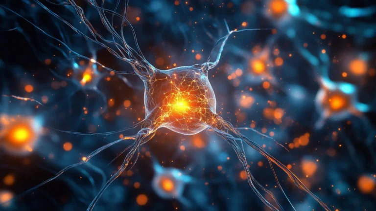
At the fundamental level, the brain’s operational capacity hinges upon synapses, the specialized junctions that facilitate the transmission of information between neurons, forming the very fabric of neural circuitry. For a considerable duration, prevailing scientific consensus posited that synaptic proliferation characterized early childhood, followed by a substantial reduction during adolescence. This established paradigm gave rise to the widely embraced theory of excessive "synaptic pruning"—a biological process involving the elimination of less efficient or redundant neural connections. The implication was that an overabundance of this pruning mechanism could potentially underlie various neuropsychiatric conditions, with schizophrenia, a disorder marked by altered perceptions of reality such as hallucinations and delusions, frequently cited as an example linked to this hypothesized over-elimination of synapses.
However, recent groundbreaking research emerging from Kyushu University has begun to dismantle this long-standing theoretical framework. A dedicated team of scientists, through their rigorous investigation, has unearthed compelling evidence that suggests the adolescent brain engages in more than just the systematic dismantling of neural connections. Their findings, detailed in a study published in the esteemed journal Science Advances on January 14th, indicate that during this developmental epoch, the brain actively establishes novel, densely clustered aggregations of synapses within specific neuronal domains.
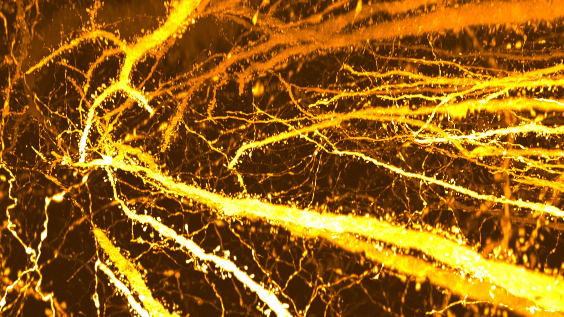
Professor Takeshi Imai, affiliated with the Faculty of Medical Sciences at Kyushu University, shared that the initial impetus for the research was not directly focused on the etiology of brain disorders. Instead, the team’s exploration was spurred by the development of an advanced, high-resolution instrument designed for synaptic analysis, introduced in 2016. Their subsequent examination of the mouse cerebral cortex, undertaken out of scientific curiosity, yielded unexpected revelations. Beyond appreciating the inherent elegance of neuronal structures, the researchers were struck by the discovery of a previously unrecognized region characterized by an exceptionally high density of dendritic spines. These spines are minute protrusions extending from dendrites, the branched extensions of nerve cells, and serve as the primary sites for the formation of excitatory synapses.
The cerebral cortex, a key area of the brain responsible for higher-level cognitive functions, is organized into six distinct layers, each contributing to the sophisticated interplay of neural circuits. Professor Imai and his collaborators specifically directed their attention to neurons residing within Layer 5. These neurons play a pivotal role in the cortical information processing hierarchy, serving as conduits that integrate signals from a multitude of sources and subsequently transmit output signals to other brain regions. Their crucial function positions them as central regulatory hubs for how the brain processes incoming and outgoing information.
To meticulously investigate the characteristics of these Layer 5 neurons, the research team employed a sophisticated combination of SeeDB2, a tissue-clearing agent developed by Imai’s group, and super-resolution microscopy. This powerful methodological approach enabled the researchers to render brain tissue optically transparent, thereby facilitating an unprecedented level of detail in mapping the distribution of dendritic spines across entire Layer 5 neurons for the first time in scientific history.
The detailed cartography of dendritic spines revealed a striking and unanticipated pattern. A distinct segment of the dendrite exhibited an unusually concentrated profusion of these spines, an aggregation the researchers have termed a "synapse hotspot." Further investigations confirmed that this specialized region is not present in the early stages of life but rather emerges and develops specifically during adolescence.
To precisely delineate the temporal emergence of this phenomenon, the researchers meticulously tracked spine distribution across various developmental stages in their mouse models. In young mice, approximately two weeks of age and prior to weaning, dendritic spines were observed to be relatively evenly dispersed across the neuronal structure. However, between the ages of three and eight weeks—a period encompassing the transition from early childhood to adolescence—a marked increase in spine density was documented within a singular region of the apical dendrite. Over time, this localized proliferation of spines culminated in the formation of a pronounced synapse hotspot. "These findings suggest that the long-established hypothesis of ‘adolescent synaptic pruning’ warrants significant re-evaluation," Professor Imai commented, underscoring the paradigm-shifting nature of their discovery.
This newfound understanding of adolescent synaptic reorganization may also offer crucial insights into the developmental pathways of certain neurological and psychiatric disorders. Ryo Egashira, the study’s lead author and a graduate student at Kyushu University’s Graduate School of Medical Sciences during the research period, posited that "while synaptic pruning occurs broadly across dendrites, synapse formation also takes place in specific dendritic compartments during adolescent cortical development. Disruption of this process may be the key factor in at least some types of schizophrenia."
To investigate this potential link, the researchers examined mouse models carrying genetic mutations in genes known to be associated with schizophrenia, including Setd1a, Hivep2, and Grin1. In these genetically modified mice, early developmental stages appeared typical, with spine density remaining within normal ranges up to two to three weeks post-birth. However, during the adolescent period, the formation of new synapses was significantly impaired, thereby preventing the proper development of the characteristic hotspot. For many years, schizophrenia has been predominantly understood as a condition characterized by an excessive loss of synapses. These recent findings introduce a compelling alternative perspective, suggesting that difficulties in constructing new synaptic connections during adolescence might play a critical, previously underappreciated role in the disorder’s pathogenesis. Nonetheless, the researchers prudently emphasize that their study was conducted exclusively on mice, and it remains an open question whether these precise developmental processes are mirrored in primate or human brains.
Looking towards the future, Professor Imai expressed the team’s ambition to identify the specific brain regions where these novel synaptic connections are being forged during adolescence. "That will tell us what circuits are actually being built during this developmental window," he stated. "Understanding how and when these connections form can advance our knowledge of both brain development and the mechanisms underlying neuropsychiatric disorders." This ongoing research holds the promise of refining our comprehension of brain plasticity during a critical developmental phase and potentially unlocking new avenues for therapeutic intervention in disorders affecting cognitive function.