Scientists at Cedars-Sinai have unveiled a groundbreaking biological mechanism that holds significant promise for developing future therapeutic interventions for spinal cord injuries, strokes, and debilitating neurological conditions such as multiple sclerosis. This pivotal research, detailed in the prestigious journal Nature, highlights an previously unrecognized but crucial function for astrocytes, a fundamental type of glial cell within the central nervous system. These cells, long understood for their supportive roles, are now revealed to be active participants in the body’s intrinsic repair processes.
The study’s senior author, neuroscientist Joshua Burda, PhD, an assistant professor of Biomedical Sciences and Neurology at Cedars-Sinai, emphasized the profound implications of these findings. "Astrocytes are essential responders when the central nervous system, encompassing the brain and spinal cord, encounters disease or injury," Dr. Burda stated. "Our investigation has demonstrated that astrocytes located at a considerable distance from the initial site of damage actively contribute to the regeneration of the spinal cord. Furthermore, we’ve elucidated a specific pathway these specialized astrocytes utilize to communicate with the immune system, directing it to efficiently clear cellular debris generated by the injury – a process absolutely vital for effective tissue restoration."
These distinct astrocytes, now termed "lesion-remote astrocytes" or LRAs, have been further categorized into several subtypes, each with unique characteristics. For the first time, this research provides a detailed explanation of how one particular LRA subtype is capable of sensing damage from afar and initiating responses that foster recovery.
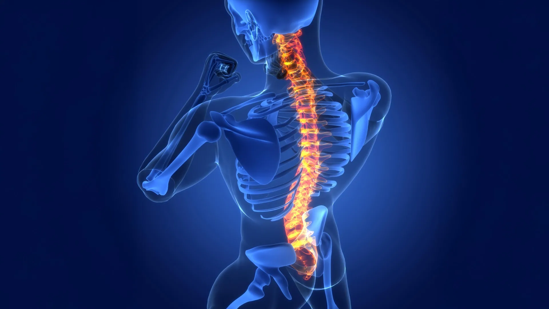
To fully appreciate the significance of these findings, it’s important to understand the intricate architecture of the spinal cord and its typical response to trauma. The spinal cord, a vital conduit extending from the brain down the body, comprises two primary regions. The inner core, known as gray matter, is densely populated with neuronal cell bodies and astrocytes. Surrounding this is the white matter, composed of astrocytes and elongated nerve fibers, or axons, which serve as the communication lines transmitting signals between the brain and the rest of the body. Astrocytes are instrumental in maintaining the precise environmental conditions required for these signals to propagate without interference.
When the spinal cord sustains an injury, such as from trauma or disease, these critical nerve fibers are severed. This disruption can lead to profound functional deficits, including paralysis and the loss of sensory perception like touch and temperature. The damaged axons fragment into cellular debris. While inflammation in most bodily tissues is typically localized to the site of injury, the extended nature of nerve fibers within the spinal cord means that damage and subsequent inflammation can propagate far beyond the initial trauma zone, complicating the healing process.
The Cedars-Sinai team’s experiments, conducted on rodent models exhibiting spinal cord injuries, provided compelling evidence that LRAs are central to promoting repair. Crucially, these observations were further validated by analyses of spinal cord tissue samples from human patients, suggesting a conserved biological mechanism across species.
A key discovery within this LRA subtype is its production of a specific protein, CCN1. This molecule acts as a signaling agent, effectively communicating with immune cells known as microglia. Dr. Burda described the role of microglia in this context: "Microglia function as the primary cleanup crew within the central nervous system. Following tissue damage, they engulf fragments of nerve fibers, which are rich in lipids. This high fat content can overwhelm their digestive capabilities, leading to a form of cellular indigestion." The research demonstrated that CCN1 secreted by astrocytes modifies microglial metabolism, enabling them to more effectively process and digest the lipid-laden debris.
This enhanced clearance of damaged cellular material, the researchers propose, may contribute to the partial, spontaneous recovery observed in some individuals following spinal cord injury. When the production of astrocyte-derived CCN1 was experimentally inhibited, the capacity for healing was significantly diminished. "Without astrocyte CCN1," Dr. Burda explained, "microglia attempt to consume the debris, but they cannot properly break it down. This leads to an accumulation of undigested material, prompting the recruitment of additional microglia, which also struggle with digestion. The result is the formation of large aggregates of debris-laden microglia, exacerbating inflammation throughout the spinal cord, which in turn impedes tissue repair."
The implications of these findings extend beyond spinal cord injuries. When researchers examined spinal cord tissue from individuals diagnosed with multiple sclerosis, they observed the same CCN1-mediated repair pathway in action. Dr. Burda noted that these fundamental principles of cellular repair may be broadly applicable to a range of injuries affecting both the brain and the spinal cord.
David Underhill, PhD, chair of the Department of Biomedical Sciences at Cedars-Sinai, underscored the underappreciated role of astrocytes in neurological healing. "The contribution of astrocytes to central nervous system repair has been remarkably under-investigated," Dr. Underhill commented. "This work strongly indicates that lesion-remote astrocytes represent a promising avenue for mitigating chronic inflammation, fostering functionally significant neural regeneration, and ultimately enhancing neurological recovery following injuries to the brain and spinal cord, as well as in various neurological diseases."
Building on this breakthrough, Dr. Burda and his team are actively pursuing the development of therapeutic strategies that can leverage the CCN1 pathway to improve outcomes for spinal cord injury patients. Their ongoing research also explores the potential influence of astrocyte CCN1 on inflammatory neurodegenerative conditions and the aging process within the nervous system.
This significant research effort involved a collaborative team of scientists. Additional authors from Cedars-Sinai include Sarah McCallum, Keshav B. Suresh, Timothy S. Islam, Manish K. Tripathi, Ann W. Saustad, Oksana Shelest, Aditya Patil, David Lee, Brandon Kwon, Katherine Leitholf, Inga Yenokian, Sophia E. Shaka, Jasmine Plummer, Vinicius F. Calsavara, and Simon R.V. Knott. Contributing authors from other institutions include Connor H. Beveridge, Palak Manchandra, Caitlin E. Randolph, Gordon P. Meares, Ranjan Dutta, Riki Kawaguchi, and Gaurav Chopra.
The research was made possible through substantial financial support from various prestigious organizations. Funding was provided by the US National Institutes of Health (NIH) through grants 5R01NS128094, R00NS105915, K99NS105915 (to J.E.B.), F31NS129372 (to K.S.), K99AG084864 (S.M.), R35 NS097303, and R01 NS123532 (RD), R01MH128866, U18TR004146, P30 CA023168, and the ASPIRE Challenge and Reduction-to-Practice award (to G.C.). Additional support came from the Paralyzed Veterans Research Foundation of America (to J.E.B.), Wings for Life (to J.E.B.), the Cedars-Sinai Center for Neuroscience and Medicine Postdoctoral Fellowship (to S.M.), the American Academy of Neurology Neuroscience Research Fellowship (to S.M.), the California Institute for Regenerative Medicine Postdoctoral Scholarship (to S.M.), the United States Department of Defense USAMRAA award W81XWH2010665 through the Peer Reviewed Alzheimer’s Research Program (to G.C.), and The Arnold O. Beckman Postdoctoral Fellowship (to C.E.R.). The Purdue University Center for Cancer Research, funded by NIH grant P30 CA023168, also contributed to this work.