
A collaborative scientific endeavor involving researchers from the German Primate Center, Hannover Medical School, and the Max Planck Institute of Molecular Cell Biology and Genetics has illuminated a fundamental mechanism underlying microcephaly, a condition characterized by an abnormally small brain size in children. This groundbreaking research, utilizing sophisticated human brain organoids cultivated in a laboratory setting, has pinpointed critical alterations in intracellular structural proteins as the culprits behind disrupted neural development.
The investigation, detailed in the scientific journal EMBO Reports, meticulously demonstrates how genetic anomalies affecting actin, a vital component of the cell’s internal scaffolding, profoundly impact the division patterns of nascent brain progenitor cells. When these foundational cells are unable to replicate and divide according to their established developmental blueprints, their population dwindles, thereby severely curtailing the overall growth trajectory of the brain and ultimately resulting in a smaller cranial volume. This discovery offers the inaugural cellular-level explanation for microcephaly observed in individuals diagnosed with the rare Baraitser-Winter syndrome.
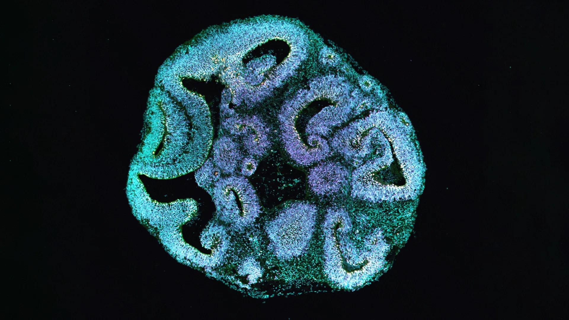
The intricate role of the cell’s internal framework, known as the cytoskeleton, in orchestrating brain development cannot be overstated. Actin, a key player within this dynamic structure, is instrumental in maintaining cellular integrity and facilitating the internal transport of molecules. In the context of Baraitser-Winter syndrome, a specific genetic mutation targets one of two critical actin-encoding genes. To meticulously investigate the ramifications of this mutation, the research team ingeniously reprogrammed skin cells obtained from affected patients, transforming them into induced pluripotent stem cells. These versatile stem cells served as the crucial starting material for the generation of three-dimensional brain organoids, effectively recapitulating the initial phases of human brain formation.
Upon observing the organoids after a developmental period of thirty days, stark divergences became apparent. Organoids derived from the cells of patients exhibited a size reduction of approximately 25 percent when contrasted with organoids cultivated from the cells of healthy donors. Furthermore, the ventricle-like compartments within these organoids, the specialized regions where progenitor cells congregate and initiate the genesis of primitive nerve cells, were notably diminished in size.
A significant shift was also detected in the composition of crucial brain cell populations. An examination of the cellular makeup within the organoids revealed a pronounced imbalance. The abundance of apical progenitor cells, which are indispensable for the construction of the cerebral cortex, was substantially lower. Concurrently, there was an observable increase in basal progenitor cells, a cell type that typically emerges later in the developmental sequence. This alteration in cell ratios strongly suggested a disruption in the temporal regulation and overall outcome of cell division processes, providing a compelling explanation for the observed failure of brain tissue to expand adequately.
The meticulous observation of how apical progenitor cells undergo division, employing high-resolution microscopy, unveiled a dramatic deviation from normal developmental pathways. Under typical circumstances, these progenitor cells predominantly divide at right angles to the ventricular surface. This precise orientation ensures the equitable distribution of cellular components and facilitates the self-renewal of apical progenitor cells, thereby maintaining a robust pool for cortical development.
However, within the organoids harboring the specific actin mutation, this cardinal division orientation underwent a significant alteration. Vertical divisions became conspicuously infrequent, with horizontal and oblique divisions becoming the predominant modes of cell replication. Consequently, the capacity of apical progenitor cells to replenish their own numbers was severely compromised. They exhibited a greater tendency to detach from the ventricular zone and were more prone to differentiate into basal progenitor cells rather than continuing their role as apical progenitors. This deviation in division orientation among progenitor cells emerges as the decisive factor precipitating the reduced brain size. It underscores the profound impact that even a single alteration within the cytoskeleton can exert on the intricate choreography of early brain development.
Further microscopic investigations using electron microscopy uncovered subtle yet significant structural anomalies at the ventricular surface. The shapes of the cells appeared irregular, and the formation of superfluous protrusions between adjacent cells was noted. Researchers also observed an unusually elevated presence of tubulin, another critical cytoskeletal protein integral to cell division, at the cell junctions. While the overarching cellular architecture remained largely intact, these minute structural imperfections may be sufficient to permanently disrupt the precise orientation of cells during division.
To definitively establish the causal link between the observed cellular defects and the specific actin mutation, the researchers conducted a pivotal control experiment. Employing the powerful CRISPR/Cas9 gene-editing technology, they introduced the identical mutation into a healthy stem cell line. The subsequent development of brain organoids derived from these genetically modified cells mirrored the precise defects observed in the organoids cultivated from patient-derived cells, unequivocally confirming the mutation as the primary driving force behind these developmental abnormalities.
This remarkable discovery offers invaluable insights into the complex interplay between rare genetic mutations and the manifestation of sophisticated brain malformations. It also powerfully demonstrates the indispensable utility of human brain organoids as a cutting-edge tool in the realm of biomedical research. The findings provide a more profound understanding of how infrequent genetic disorders can lead to intricate disruptions in brain architecture and simultaneously highlight the immense potential of brain organoids for advancing our knowledge in this field.
The therapeutic implications of this study are primarily centered on enhanced diagnostics. The data generated significantly aids in the more precise classification of genetic findings in patients presenting with microcephaly. Given that the condition impacts fundamental processes during early fetal development, direct therapeutic interventions in humans present considerable complexities. Nevertheless, the exploration of novel pharmacological agents designed to modulate the intricate interactions between actin and microtubules holds promise for opening up new avenues for treatment in the long term. This research not only unravels a fundamental biological process but also paves the way for future diagnostic and therapeutic advancements in the study of developmental brain disorders.
About the Author


