
At the heart of this innovation lies a sophisticated imaging apparatus designed to meticulously detect exceptionally faint signals emanating from surface-enhanced Raman scattering (SERS) nanoparticles. These specially designed nanoparticles are engineered to selectively adhere to biomarkers indicative of cancerous cells. Once these probes are introduced to a biological sample or a region under examination, the imaging system meticulously analyzes their characteristic Raman signatures, automatically pinpointing areas exhibiting a higher probability of containing malignant tissue.
Dr. Zhen Qiu, the principal investigator of the research team and a leader at Michigan State University’s Institute for Quantitative Health Science and Engineering (IQ), highlighted the limitations of current diagnostic methodologies. "Conventional approaches to cancer-related diagnoses are often protracted and resource-intensive," Dr. Qiu explained. "They necessitate the staining of tissue samples and the meticulous scrutiny of pathologists to identify any anomalies." While acknowledging that this novel system would not immediately supplant the definitive role of pathology, Dr. Qiu emphasized its potential as a swift screening mechanism to expedite the diagnostic process.
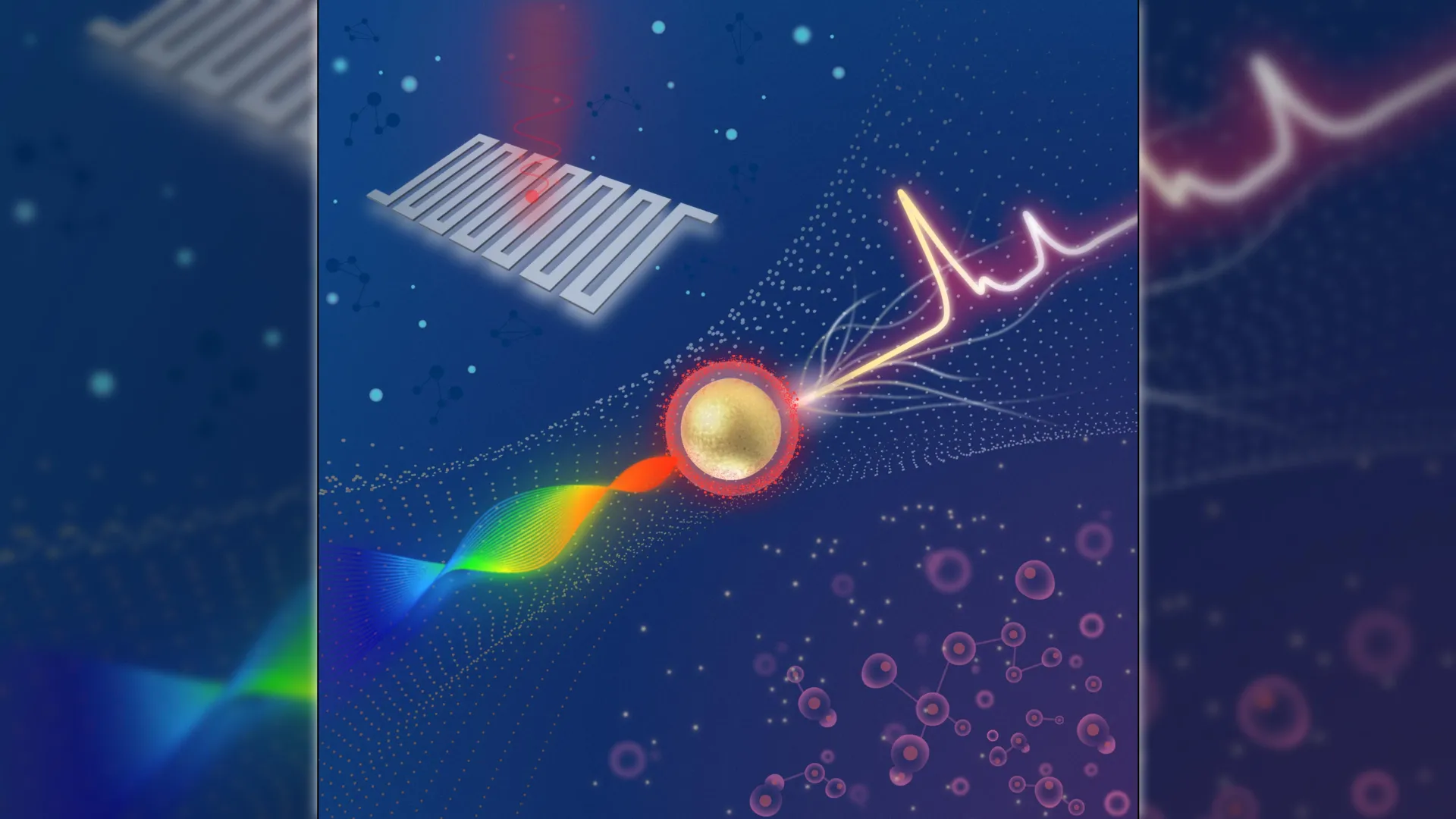
The efficacy of this advanced system has been underscored by published findings, which reveal substantial enhancements in detection sensitivity. In a recent publication in Optica, a journal renowned for high-impact research from Optica Publishing Group, Dr. Qiu and his collaborators detailed how their system can differentiate between cancerous and healthy cells by detecting Raman signals that are approximately four times weaker than those typically measured by comparable commercially available systems. This significant leap in sensitivity is attributed to a synergistic combination of a swept-source laser, which dynamically alters its wavelength during the analysis, and an extraordinarily sensitive detector known as a superconducting nanowire single-photon detector (SNSPD).
"The potential applications of this technology are vast," Dr. Qiu elaborated. "It could ultimately lead to the development of portable or intraoperative devices that empower clinicians to detect cancers at their earliest stages, enhance the precision of biopsy sampling, and monitor disease progression through less invasive testing." He further posited that such advancements could profoundly improve patient outcomes by minimizing diagnostic delays, thereby accelerating the crucial transition from detection to timely treatment initiation.
The pursuit of pushing the boundaries of detection limits is significantly bolstered by the integration of superconducting detectors. Dr. Qiu’s research group has been actively investigating the utility of SNSPDs in augmenting a diverse array of imaging technologies. These detectors operate on the principle of a superconducting wire capable of registering individual photons, thereby enabling the system to capture exceedingly faint optical signals with remarkable speed while simultaneously minimizing extraneous background noise.
For this particular project, the researchers set out to construct a platform that could measure Raman signals far more attenuated than those discernible by existing Raman systems. Raman imaging functions by generating a detailed map of a sample’s chemical composition by analyzing the unique light-scattering patterns, or "fingerprints," of its constituent molecules. The intensity of these intrinsic signals can be amplified through the strategic application of SERS nanoparticles.
"The integration of this cutting-edge detector with a swept-source Raman architecture, which ingeniously replaces a cumbersome camera and achieves more efficient light collection, has resulted in a system exhibiting a detection threshold significantly surpassing that of comparable commercial systems," Dr. Qiu stated. He also noted that the fiber-optic coupling configuration and the compact design of the system are instrumental in facilitating miniaturization and expediting its eventual translation into clinical settings.
The system has demonstrated robust tumor contrast across a variety of sample types. To rigorously evaluate its performance, the research team employed SERS nanoparticles functionalized with hyaluronan acid. This specific coating facilitates the nanoparticles’ binding to CD44, a surface protein frequently overexpressed on numerous types of tumor cells. Initial experimental trials, conducted using straightforward nanoparticle solutions, indicated that the system could achieve an impressive femtomolar level of sensitivity. Subsequently, the imaging platform was applied to investigate cultured breast cancer cells, excised mouse tumors, and samples of healthy tissue.
"The SERS signals were observed to be highly concentrated within the tumor samples, with only minimal background signals detected in the healthy tissue," Dr. Qiu reported. "This finding serves as a compelling demonstration of both the system’s extraordinary sensitivity and its capacity to deliver reliable discrimination between tumor and healthy tissues." He further elaborated that by modifying or substituting the targeting molecule, this innovative methodology could be readily adapted to detect a wide spectrum of other cancer types.
Looking ahead, the path toward clinical implementation necessitates further research and development. According to the researchers, additional work is essential before the system can be reliably deployed in clinical environments. Future enhancements will primarily focus on augmenting the readout speed of the system and broadening the scope of validation studies. The team is actively exploring the integration of faster laser sources, including vertical-cavity surface-emitting lasers (VCSELs), and is investigating whether constricting the wavelength sweep range could yield further performance improvements. Moreover, they are planning multiplexing experiments, which would involve the simultaneous use of different nanoparticles to target multiple biomarkers, thereby providing a more comprehensive diagnostic profile. The researchers expressed their gratitude to industry collaborator Quantum Opus for providing the SNSPD devices that were integral to this groundbreaking work.
About the Author


