
A groundbreaking discovery originating from Cedars-Sinai has illuminated a previously unrecognized biological repair mechanism, offering significant promise for the development of novel therapeutic strategies targeting spinal cord injuries, strokes, and neurodegenerative conditions like multiple sclerosis. This pivotal research, detailed in the esteemed scientific journal Nature, reveals an unexpected and crucial role for astrocytes, a fundamental class of glial cells within the central nervous system. These cells, long understood as essential support structures for neurons, have now been shown to actively participate in healing processes, even when situated far from the site of damage.
"Astrocytes are fundamental players in the body’s response to insults and disorders affecting the central nervous system, encompassing both the brain and the spinal cord," explained Joshua Burda, PhD, a neuroscientist and assistant professor of Biomedical Sciences and Neurology at Cedars-Sinai, who served as the senior author of the study. "Our investigation has unveiled that astrocytes located at a considerable distance from an injury site actually contribute significantly to the restorative processes of the spinal cord. Furthermore, our work has elucidated a specific pathway through which these specialized astrocytes orchestrate the immune system’s response to effectively clear cellular debris generated by the injury, a process that is absolutely vital for tissue regeneration."
The research team has formally designated these remote-acting astrocytes as "lesion-remote astrocytes," or LRAs. Within this newly identified category, they have further distinguished several distinct subtypes, each with unique functional characteristics. For the first time, this study provides a comprehensive explanation of how one particular LRA subtype possesses the remarkable ability to sense damage from afar and initiate responses that actively promote functional recovery.
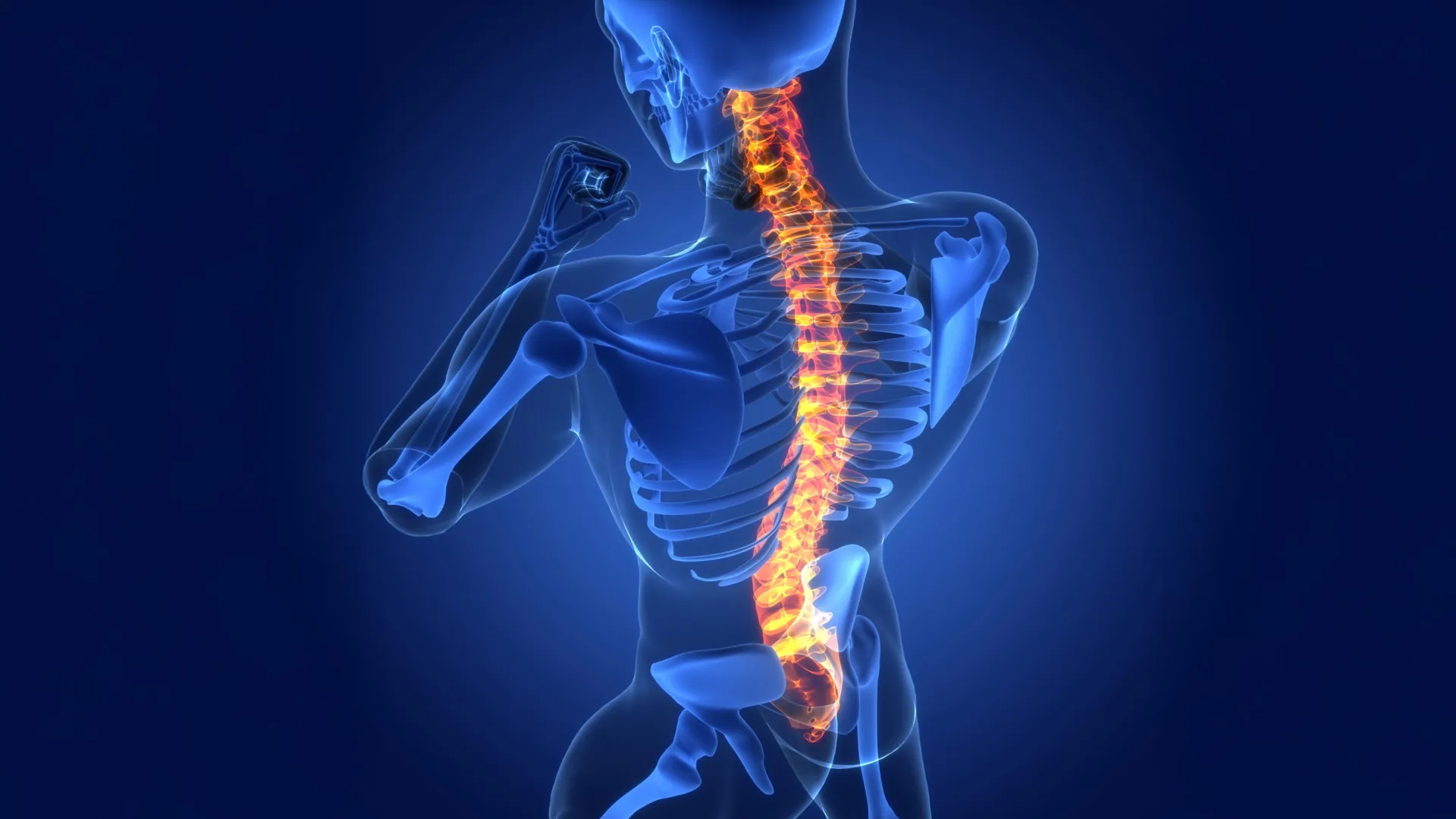
Understanding the intricate response of the spinal cord to injury is crucial for appreciating the significance of this discovery. The spinal cord itself is an extensive conduit of neural tissue extending from the brain down the vertebral column. Internally, it features a region known as gray matter, which is densely populated with nerve cell bodies and astrocytes. This is enveloped by white matter, primarily composed of astrocytes and the long axons of nerve fibers that transmit vital electrochemical signals between the brain and the rest of the body. The primary function of astrocytes in this context is to meticulously maintain a stable microenvironment, ensuring the unimpeded and efficient propagation of these neural signals.
When the spinal cord sustains an injury, such as from trauma or disease, these delicate nerve fibers are severed, leading to a cascade of detrimental effects. This physical disruption can manifest as paralysis, loss of motor control, and a profound alteration or complete loss of sensory perception, including touch, temperature, and pain. The severed nerve fibers subsequently degrade, breaking down into cellular fragments and molecular debris. In most bodily tissues, the inflammatory response to injury is typically localized to the affected area. However, the unique anatomical structure of the spinal cord, with its nerve fibers extending considerable distances, means that damage and subsequent inflammation can readily spread far beyond the initial point of insult, exacerbating the overall injury.
The investigation into lesion-remote astrocytes and their role in immune-mediated cleanup has yielded compelling insights. Through meticulous experiments conducted on rodent models afflicted with spinal cord injuries, the researchers observed that LRAs are instrumental in fostering the repair process. Crucially, their findings also revealed robust indicators suggesting that this same biological mechanism is operative within human spinal cord tissue obtained from patients.
A key finding centers on a specific subtype of LRA that synthesizes a protein known as CCN1. This molecule acts as a critical signaling agent, transmitting instructions to resident immune cells within the central nervous system, specifically microglia.
"Microglia are essentially the primary ‘waste management’ crew of the central nervous system," elaborated Dr. Burda, drawing an analogy to clarify their function. "Following tissue damage, their primary role is to engulf and clear away fragments of nerve fibers. This debris is particularly rich in fats, which can present a digestive challenge for microglia, leading to what might be described as cellular indigestion. Our experimental results demonstrated that the CCN1 produced by astrocytes effectively signals these microglia, prompting them to alter their metabolic processes. This metabolic shift enables them to more efficiently process and digest the fatty components of the nerve fiber debris."
Dr. Burda further posited that this enhanced clearance of cellular debris could potentially explain why certain individuals experience partial, spontaneous recovery following spinal cord injuries. Conversely, when the researchers experimentally inhibited or eliminated the production of astrocyte-derived CCN1, the healing process was observed to be significantly impaired.
"When we disable the astrocyte’s ability to produce CCN1, the microglia still attempt to ingest the debris, but they are unable to properly digest it," Dr. Burda explained. "This leads to a situation where they accumulate undigested material and then signal for additional microglia to be recruited, which also ingest but cannot effectively digest. This results in the formation of substantial agglomerations of microglia engorged with debris, intensifying inflammation throughout the spinal cord. Consequently, the damaged tissue is unable to repair itself as effectively."
The implications of this discovery extend beyond spinal cord injuries, holding potential relevance for other neurological conditions. When scientists meticulously examined spinal cord samples from individuals diagnosed with multiple sclerosis, they observed the presence of the same CCN1-mediated repair process. Dr. Burda highlighted that these fundamental principles of neural repair might be broadly applicable to a range of injuries affecting either the brain or the spinal cord.
"The contribution of astrocytes to the healing processes within the central nervous system has been remarkably under-explored until now," commented David Underhill, PhD, chair of the Department of Biomedical Sciences at Cedars-Sinai. "This seminal work strongly indicates that lesion-remote astrocytes represent a highly promising avenue for mitigating chronic inflammation, promoting functionally meaningful neural regeneration, and ultimately enhancing neurological recovery following injuries to the brain and spinal cord, as well as in the context of various neurological diseases."
Building upon these foundational findings, Dr. Burda is now actively engaged in developing therapeutic strategies designed to leverage the CCN1 signaling pathway to improve spinal cord healing outcomes. His research group is also investigating the potential role of astrocyte CCN1 in modulating inflammatory neurodegenerative diseases and the aging process.
The study involved a comprehensive team of researchers from Cedars-Sinai, including Sarah McCallum, Keshav B. Suresh, Timothy S. Islam, Manish K. Tripathi, Ann W. Saustad, Oksana Shelest, Aditya Patil, David Lee, Brandon Kwon, Katherine Leitholf, Inga Yenokian, Sophia E. Shaka, Jasmine Plummer, Vinicius F. Calsavara, and Simon R.V. Knott. Additional contributions were made by Connor H. Beveridge, Palak Manchandra, Caitlin E. Randolph, Gordon P. Meares, Ranjan Dutta, Riki Kawaguchi, and Gaurav Chopra.
The research was generously supported by several prestigious funding bodies, including the US National Institutes of Health (NIH) through grants 5R01NS128094, R00NS105915, K99NS105915 (to J.E.B.), F31NS129372 (to K.S.), K99AG084864 (S.M.), R35 NS097303 and R01 NS123532 (RD), R01MH128866, U18TR004146, P30 CA023168, and the ASPIRE Challenge and Reduction-to-Practice award (to G.C.). Further support was provided by the Paralyzed Veterans Research Foundation of America (to J.E.B.), Wings for Life (to J.E.B.), the Cedars-Sinai Center for Neuroscience and Medicine Postdoctoral Fellowship (to S.M.), the American Academy of Neurology Neuroscience Research Fellowship (to S.M.), the California Institute for Regenerative Medicine Postdoctoral Scholarship (to S.M.), the United States Department of Defense USAMRAA award W81XWH2010665 through the Peer Reviewed Alzheimer’s Research Program (to G.C.), The Arnold O. Beckman Postdoctoral Fellowship (to C.E.R.), and the Purdue University Center for Cancer Research, funded by NIH grant P30 CA023168.
About the Author


