
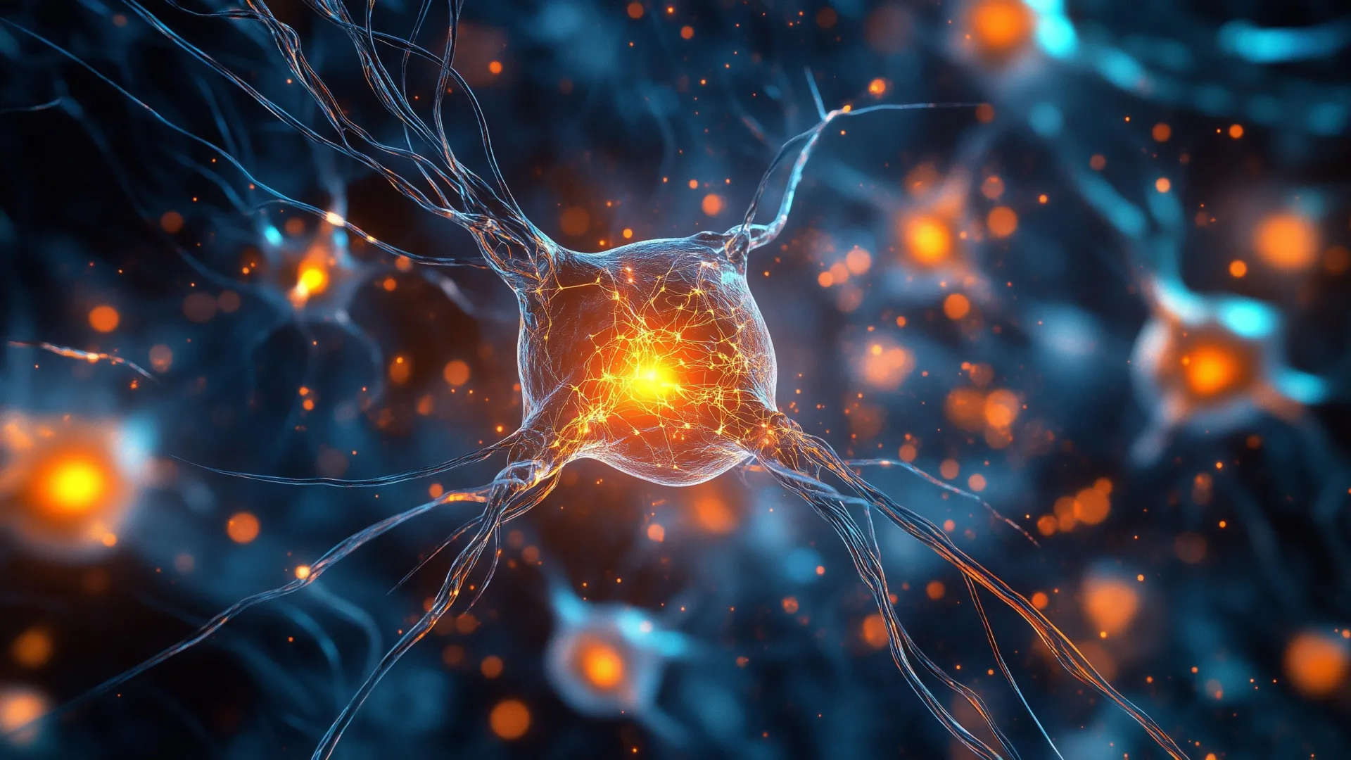
A pioneering "zap-and-freeze" technique, developed by researchers at Johns Hopkins Medicine, has achieved a significant milestone by capturing the lightning-fast exchange of information between brain cells in living tissue, a feat previously beyond the reach of scientific observation. This innovative approach allows scientists to witness neural interactions as they happen, providing unprecedented insight into the intricate workings of the brain. The groundbreaking findings, detailed in the November 24th edition of the journal Neuron and bolstered by support from the National Institutes of Health, hold immense promise for unraveling the biological underpinnings of non-hereditary forms of Parkinson’s disease.
Parkinson’s disease, a progressive neurodegenerative disorder, manifests in various forms, with the majority of cases being sporadic and not directly inherited. These sporadic instances are characterized by disruptions at the synapse, the minuscule junction where one nerve cell transmits signals to another. The inherent smallness of this crucial communication hub and the sheer speed at which its processes unfold have long presented formidable challenges to detailed scientific study. Dr. Shigeki Watanabe, an associate professor of cell biology at Johns Hopkins Medicine and the senior author of the current study, emphasized the historical difficulty in observing these rapid synaptic events.
The research team’s hope is that this novel method for visualizing synaptic membrane dynamics in live brain tissue will illuminate both the shared and distinct characteristics between inherited and non-inherited variations of Parkinson’s disease. Dr. Watanabe further suggested that the technique’s capabilities could eventually pave the way for the development of targeted therapeutic interventions for this debilitating neurological condition.
At the heart of a healthy brain’s function lies a sophisticated communication network, reliant on the precise delivery of chemical messages between neurons. This crucial transmission is facilitated by synaptic vesicles, minute sacs that encapsulate and transport these vital chemical messengers. The seamless operation of this vesicular system is fundamental to essential cognitive processes such as learning, memory formation, and the overall processing of information. Dr. Watanabe underscored that a thorough understanding of normal vesicle behavior is paramount to identifying the precise points at which neural communication begins to falter in the context of neurological diseases.
The "zap-and-freeze" methodology itself is an evolution of Dr. Watanabe’s prior work. He was instrumental in designing an earlier iteration of this approach, which was detailed in a 2020 publication in Nature Neuroscience. That foundational technique employed a brief but potent electrical stimulus to instantaneously activate brain tissue, immediately followed by a rapid freezing process. This swift action effectively arrests cellular structures in their exact configurations, preserving them for subsequent, high-resolution examination using electron microscopy. This preservation of transient states is what allows for the visualization of phenomena that occur in fractions of a second.
In a related study published earlier this year in Nature Neuroscience, Dr. Watanabe applied this advanced imaging technique to genetically modified mice. The focus of that investigation was to explore the role of a specific protein, known as intersectin. The findings from that research elucidated how intersectin plays a critical role in tethering synaptic vesicles to a precise location within the neuron, ensuring they remain poised for release until the opportune moment to transmit their signal to an adjacent neuron.
For the latest research, the Johns Hopkins team extended their application of the "zap-and-freeze" technique to human brain tissue. They meticulously examined samples from healthy laboratory mice and conducted a comparative analysis with living cortical brain tissue. This human tissue was ethically obtained, with explicit consent, from six individuals undergoing epilepsy surgery at The Johns Hopkins Hospital. These surgical procedures were necessitated by the presence of hippocampal lesions, highlighting the critical need for advanced research tools to understand conditions affecting such vital brain structures.
In collaboration with esteemed colleagues Jens Eilers and Kristina Lippmann from Leipzig University in Germany, the researchers first rigorously validated the efficacy of the "zap-and-freeze" method within mouse brain tissue. Their initial confirmation involved the precise observation of calcium signaling, a well-established trigger that initiates the release of neurotransmitters from neurons. This step was crucial to ensure the technique’s reliability and accuracy in capturing fundamental neural events.
Subsequently, the team utilized the established method to stimulate mouse neurons and meticulously documented the pivotal moment when synaptic vesicles merge with the neuronal cell membrane, thereby releasing their chemical cargo. Beyond this release phase, the researchers also captured and analyzed the subsequent processes by which the cells efficiently retrieve and recycle these vesicles. This critical cellular mechanism is known as endocytosis.
Remarkably, when the "zap-and-freeze" technique was applied to the human brain tissue samples, the researchers observed the identical endocytotic vesicle recycling steps occurring within human neurons. This finding strongly suggests a conserved fundamental process across species.
A key protein, identified as Dynamin1xA, was found to be present in both mouse and human brains at the specific sites where endocytosis is believed to occur. This protein is essential for the rapid recycling of synaptic membranes. The identification of this conserved protein reinforces the notion that the molecular mechanisms governing ultrafast endocytosis are remarkably similar in both mouse and human neural tissues. Dr. Watanabe highlighted this conservation, stating that it significantly strengthens the scientific rationale for employing mouse models in the study of human brain biology.
Looking towards future applications, Dr. Watanabe expressed his keen interest in applying the "zap-and-freeze" method to brain tissue collected from individuals diagnosed with Parkinson’s disease who are undergoing deep brain stimulation procedures. The ultimate objective of this future research would be to directly observe and characterize any potential alterations in vesicle dynamics within neurons affected by the disease. Such insights could be invaluable in understanding the pathological cascade of Parkinson’s and potentially identifying novel therapeutic targets.
The research initiative was made possible through substantial funding from a consortium of esteemed organizations, including the National Institutes of Health (under grants U19 AG072643, 1DP2 NS111133-01, 1R01 NS105810-01A1, R35 NS132153, and S10RR026445), the Howard Hughes Medical Institute, the Kazato Foundation, the American Lebanese Syrian Associated Charities, the Marine Biological Laboratory, Leipzig University, the Roland Ernst Stiftung, Johns Hopkins Medicine, the Chan Zuckerberg Initiative, the Brain Research Foundation, the Helis Foundation, the Robert J Kleberg Jr and Helen C Kleberg Foundation, the McKnight Foundation, the Esther A. & Joseph Klingenstein Fund, and the Vallee Foundation.
The collaborative effort behind this significant advancement involved a dedicated team of researchers. From Johns Hopkins, contributors included Chelsy Eddings, Minghua Fan, Yuuta Imoto, Kie Itoh, Xiomara McDonald, William Anderson, Paul Worley, and David Nauen. They worked in close partnership with Jens Eilers and Kristina Lippmann from Leipzig University, underscoring the international nature of cutting-edge scientific discovery.
About the Author


