
Researchers at Cedars-Sinai have illuminated a previously unrecognized biological cascade that holds significant promise for developing novel therapeutic strategies targeting debilitating conditions like spinal cord injuries, strokes, and neurodegenerative disorders such as multiple sclerosis. This groundbreaking discovery, detailed in the esteemed scientific journal Nature, highlights an unexpected and pivotal role played by astrocytes, a fundamental class of glial cells integral to the central nervous system. These specialized cells, situated far from the immediate site of damage, are now understood to be active participants in the intricate process of tissue regeneration.
"Astrocytes are not merely passive bystanders; they are crucial architects of the response to pathology and injury within the central nervous system, encompassing both the brain and the spinal cord," stated neuroscientist Joshua Burda, PhD, an assistant professor of Biomedical Sciences and Neurology at Cedars-Sinai and the senior author of the study. "Our investigation revealed that astrocytes located at a considerable distance from an injury site actively contribute to the restorative processes within the spinal cord. Furthermore, we elucidated a sophisticated mechanism by which these unique astrocytes engage the immune system, directing it to effectively clear cellular debris generated by the injury, a step that is absolutely essential for successful tissue repair."
The research team has designated these newly identified cells as "lesion-remote astrocytes," or LRAs, and has further categorized them into several distinct subtypes. This work represents the first comprehensive explanation of how one particular LRA subtype possesses the remarkable ability to perceive damage from afar and initiate a cascade of responses that are conducive to functional recovery.
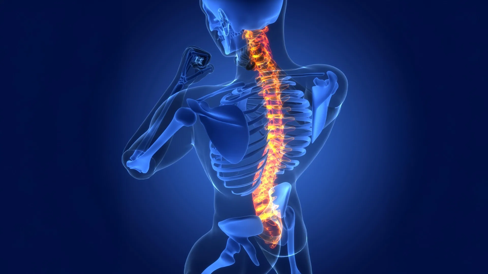
To fully appreciate the significance of LRAs, it is important to understand the fundamental architecture and response of the spinal cord to trauma. The spinal cord itself is a complex, elongated column of neural tissue extending from the brain down the vertebral column. Internally, it is comprised of gray matter, which houses the cell bodies of neurons alongside astrocytes, and is enveloped by white matter, characterized by myelinated nerve fibers—axons—that are also supported by astrocytes. The primary function of astrocytes in their typical role is to meticulously maintain a stable and optimal microenvironment, ensuring the unimpeded transmission of electrical and chemical signals between the brain and the peripheral nervous system.
Upon experiencing injury, such as a contusion or laceration, the delicate network of nerve fibers within the spinal cord becomes severed. This disruption can lead to profound motor deficits, resulting in paralysis, and sensory impairments, affecting sensations like touch, pain, and temperature. The physical tearing of these fibers results in the fragmentation of cellular components, creating a significant amount of cellular debris. While inflammation in most tissues is typically localized to the immediate area of injury, the extensive longitudinal reach of nerve fibers within the spinal cord means that the consequences of damage and subsequent inflammation can propagate far beyond the initial point of impact, affecting a broader expanse of neural tissue.
The Cedars-Sinai study, employing experimental models involving mice subjected to spinal cord injuries, provided compelling evidence for the critical role LRAs play in facilitating repair. These findings were further corroborated by observations in spinal cord tissue samples obtained from human patients, indicating a conserved biological mechanism across species.
A key discovery within this LRA subtype is its production of a specific protein known as CCN1. This signaling molecule acts as a crucial intermediary, communicating with resident immune cells in the central nervous system, specifically microglia. Microglia are the primary immune surveillance and phagocytic cells of the brain and spinal cord.
"One of the principal responsibilities of microglia is to act as the chief ‘sanitation crew’ within the central nervous system," Dr. Burda explained. "Following tissue damage, they are tasked with engulfing fragments of severed nerve fibers. These fragments are rich in lipids, which can overwhelm the microglia’s metabolic capacity, leading to a state akin to indigestion. Our experimental data clearly demonstrated that the CCN1 protein produced by astrocytes signals to these microglia, prompting a metabolic shift that enhances their ability to efficiently process and digest these fatty cellular remnants."
Dr. Burda further elaborated that this enhanced debris clearance mechanism may offer a biological explanation for why some individuals experience a degree of spontaneous functional recovery following spinal cord injury. Crucially, when the researchers experimentally inhibited the production of astrocyte-derived CCN1, the observed healing processes were significantly impaired.
"In situations where astrocyte CCN1 is absent, the microglia are still able to engulf debris, but their capacity to digest it is severely compromised," Dr. Burda noted. "This leads to a build-up of undigested material within the microglia, which in turn triggers an amplified recruitment of more microglia. These newly arrived cells also engage in the same inefficient process, resulting in the formation of large aggregates of debris-laden microglia. This accumulation exacerbates inflammation throughout the spinal cord, ultimately hindering the tissue’s ability to repair effectively."
The implications of this discovery extend beyond spinal cord injuries, offering potential insights into other neurological conditions. When scientists examined spinal cord tissue from individuals diagnosed with multiple sclerosis, they observed evidence of the same CCN1-mediated repair process. Dr. Burda suggested that these fundamental principles of neural repair may be broadly applicable to a range of injuries and diseases affecting both the brain and the spinal cord.
"The multifaceted role of astrocytes in the complex landscape of central nervous system healing has been remarkably underappreciated until now," commented David Underhill, PhD, chair of the Department of Biomedical Sciences at Cedars-Sinai. "This pioneering research strongly indicates that lesion-remote astrocytes represent a highly promising therapeutic target for mitigating chronic inflammation, fostering functionally meaningful regeneration, and ultimately promoting neurological recovery in the aftermath of brain and spinal cord injuries, as well as in the context of various neurological diseases."
Building on these pivotal findings, Dr. Burda and his team are actively engaged in developing innovative therapeutic strategies that aim to leverage the CCN1 signaling pathway to enhance spinal cord healing. Their ongoing research also seeks to elucidate how astrocyte CCN1 might influence the progression of inflammatory neurodegenerative diseases and the aging process within the nervous system.
The Cedars-Sinai research team involved in this study includes Sarah McCallum, Keshav B. Suresh, Timothy S. Islam, Manish K. Tripathi, Ann W. Saustad, Oksana Shelest, Aditya Patil, David Lee, Brandon Kwon, Katherine Leitholf, Inga Yenokian, Sophia E. Shaka, Jasmine Plummer, Vinicius F. Calsavara, and Simon R.V. Knott. Additional contributions were made by Connor H. Beveridge, Palak Manchandra, Caitlin E. Randolph, Gordon P. Meares, Ranjan Dutta, Riki Kawaguchi, and Gaurav Chopra.
This research was made possible through substantial financial support from various esteemed organizations. Key funding was provided by the US National Institutes of Health (NIH) through grants 5R01NS128094, R00NS105915, and K99NS105915 (to J.E.B.), F31NS129372 (to K.S.), K99AG084864 (S.M.), R35 NS097303 and R01 NS123532 (RD), R01MH128866, U18TR004146, P30 CA023168, and the ASPIRE Challenge and Reduction-to-Practice award (to G.C.). Further support was received from the Paralyzed Veterans Research Foundation of America (to J.E.B.), Wings for Life (to J.E.B.), the Cedars-Sinai Center for Neuroscience and Medicine Postdoctoral Fellowship (to S.M.), the American Academy of Neurology Neuroscience Research Fellowship (to S.M.), and the California Institute for Regenerative Medicine Postdoctoral Scholarship (to S.M.). Additional funding came from the United States Department of Defense USAMRAA award W81XWH2010665 through the Peer Reviewed Alzheimer’s Research Program (to G.C.), and The Arnold O. Beckman Postdoctoral Fellowship (to C.E.R.). The Purdue University Center for Cancer Research, funded by NIH grant P30 CA023168, also contributed to this work.
About the Author


